组成图示
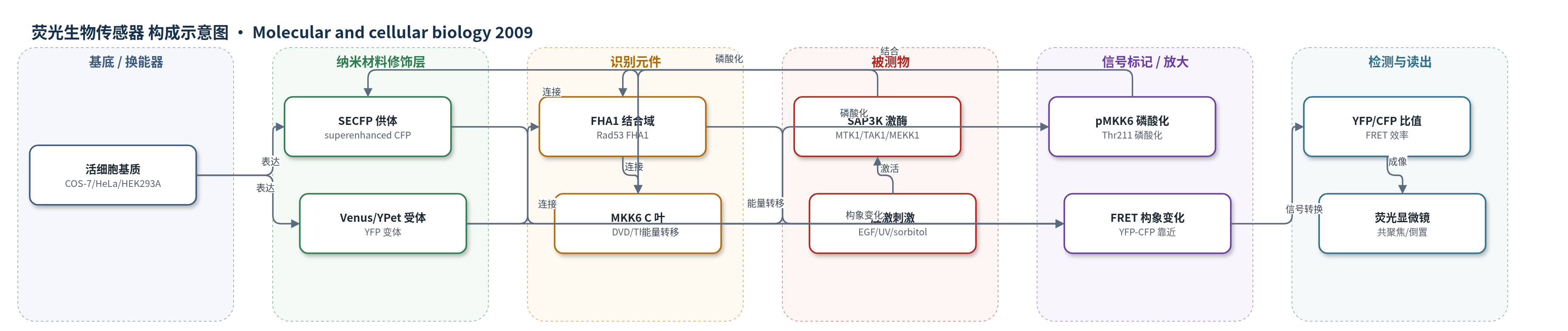
点击图片查看大图 · 依据论文自动绘制
传感器类型
荧光生物传感器
检测对象
SAP3K 激酶活性(stress-activated protein kinase kinase kinases, SAP3Ks;MTK1/MEKK4、MEKK1、TAK1、ASK1、TAO2);样品基质:活细胞(COS-7、HeLa、HEK293A),受 EGF、TNF-α、sorbitol 渗透压、anisomycin、UV-C、MMS、etoposide 刺激
检测原理
该传感器为基因编码 FRET 报告蛋白。活细胞中,应激刺激激活 SAP3K 激酶(如 MTK1、TAK1、MEKK1)。SAP3K 通过 MKK6 C 端叶的 DVD docking 位点识别并磷酸化 MKK6 的 Thr-211/Ser-207。磷酸化 Thr-211 被 Rad53 FHA1 结构域结合,使 FHA1 与 MKK6 发生分子内相互作用,拉近 N 端 YFP(Venus/YPet)与 C 端 SECFP 的距离,提高 FRET 效率。405/440 nm 激发下,YFP 发射增强、CFP 发射减弱,YFP/CFP 比值升高,从而反映 SAP3K 活性。内源磷酸酶去磷酸化可使信号恢复,实现可逆监测;NLS 或 CAAX 序列可将传感器定位到细胞核或质膜,用于区室化检测。
检测灵敏度
—
效应效果
L225/YL225 选择性响应能磷酸化 MKK6 的 SAP3Ks(MTK1、MEKK1、TAK1、ASK1、TAO2),不响应 Raf-1;全长 MTK1/TAK1 需 GADD45α/TAB1 激活。YFP/CFP 比值随 MTK1N 剂量增加,与免疫印迹磷酸化一致。TNF-α、IL-1β、anisomycin、MMS、UV-C、sorbitol、EGF 均可诱导信号,IL-1β 洗脱后恢复。稳定表达 YL225 的 HEK293A 中,sorbitol/anisomycin 诱导的内源 MKK3/6、MKK4、p38、JNK 激活与亲本细胞相似。anisomycin 半数激活时间 20.8±2.7 min,MMS 为 97.4±29.5 min;EGF/渗透压定位于质膜,anisomycin/UV-C 定位于细胞质,etoposide 定位于细胞核。可用于高通量筛选 SAPK 调节剂。
传感器的构成
- 表达基质:COS-7、HeLa 或 HEK293A 细胞,承载报告蛋白并接收 EGF、TNF-α、sorbitol、anisomycin、UV-C、MMS、etoposide 等刺激
- 荧光受体:Venus 或 YPet(YFP 变体),位于 N 端,作为 FRET 受体
- 连接肽:linker 1(LD)、linker 2(VE)、linker 3(GGRGG),连接各结构域并调节构象
- 磷酸化识别元件:Rad53 FHA1 结构域(aa22-162),结合磷酸化 Thr-211 肽段,介导构象变化
- 激酶底物/识别元件:人 MKK6 C 端叶(aa132-334),含 Ser-207/Thr-211 磷酸化位点和 DVD docking 位点,被 SAP3K 磷酸化
- 荧光供体:SECFP(superenhanced CFP),位于 C 端,作为 FRET 供体
- 亚细胞定位序列:双 NLS 或 K-Ras CAAX 膜定位序列,将报告器导向细胞核或质膜
- 优化突变:MKK6 激活环 D213A、A214D 和 P218 缺失,增强 FHA1 识别与 FRET 响应
- 信号读出:YFP/CFP 发射比(405/440 nm 激发,525-555/455-510 nm 发射),由共聚焦或倒置荧光显微镜读取
中文摘要
应激激活蛋白激酶(SAPKs,包括 p38 和 JNK)是 MAPK 家族成员,在细胞暴露于紫外线、渗透压等环境压力时决定细胞命运。SAPKs 由 SAP2Ks 激活,SAP2Ks 又由多种 SAP3Ks 激活。传统磷酸化特异性抗体免疫印迹只能测量细胞群体中 SAP3K 的平均活性,难以揭示其细胞内动态。本文开发了一种基于荧光共振能量转移(FRET)的 MKK6 SAP2K 活性报告器,可在完整单细胞中实时显示 SAP3K 激活行为。利用该报告器,作者证明 SAP3K 激活在细胞群体中可同步或异步发生,并在单细胞不同区室中发生,取决于所受压力类型。具体而言,表皮生长因子和渗透压主要在质膜激活 SAP3Ks,anisomycin 和紫外线主要在细胞质激活,etoposide 主要在细胞核激活。这些结果揭示了 SAPK 反应中此前未知的异质性,并回答了不同压力诱导刺激特异性 SAPK 反应的细胞位置问题。
英文摘要
The stress-activated protein kinases (SAPKs), namely, p38 and JNK, are members of the mitogen-activated protein kinase family and are important determinants of cell fate when cells are exposed to environmental stresses such as UV and osmostress. SAPKs are activated by SAPK kinases (SAP2Ks), which are in turn activated by various SAP2K kinases (SAP3Ks). Because conventional methods, such as immunoblotting using phospho-specific antibodies, measure the average activity of SAP3Ks in a cell population, the intracellular dynamics of SAP3K activity are largely unknown. Here, we developed a reporter of SAP3K activity toward the MKK6 SAP2K, based on fluorescence resonance energy transfer, that can uncover the dynamic behavior of SAP3K activation in cells. Using this reporter, we demonstrated that SAP3K activation occurs either synchronously or asynchronously among a cell population and in different cellular compartments in single cells, depending on the type of stress applied. In particular, SAP3Ks are activated by epidermal growth factor and osmostress on the plasma membrane, by anisomycin and UV in the cytoplasm, and by etoposide in the nucleus. These observations revealed previously unknown heterogeneity in SAPK responses and supplied answers to the question of the cellular location in which various stresses induce stimulus-specific SAPK responses.