组成图示
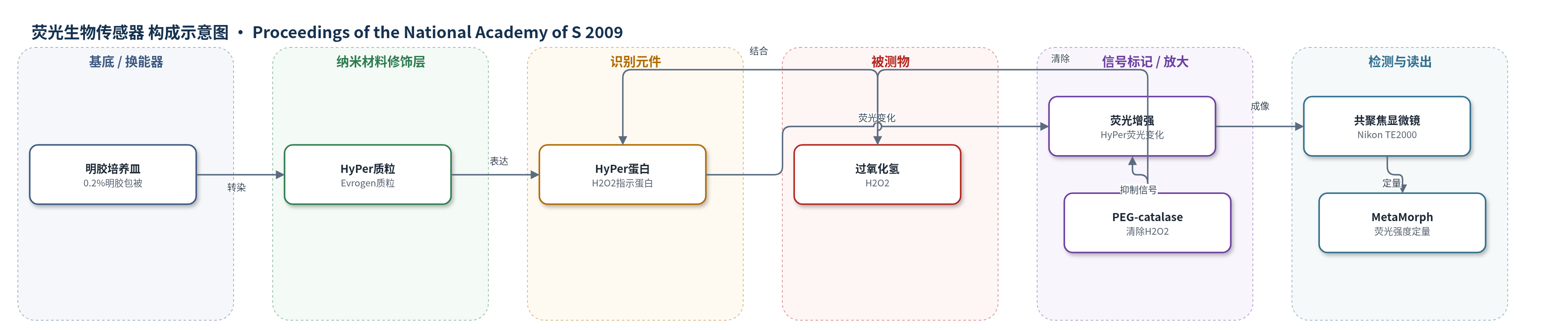
点击图片查看大图 · 依据论文自动绘制
传感器类型
荧光生物传感器
检测对象
过氧化氢(H2O2);样品基质:培养牛主动脉内皮细胞(BAECs)胞内、eNOS 敲低/抑制细胞、高糖培养细胞
检测原理
HyPer 是一种基因编码的胞质靶向荧光 H2O2 指示蛋白。将 HyPer 质粒转染 BAECs 后,细胞表达 HyPer 蛋白。当胞内 H2O2 浓度升高时,H2O2 氧化 HyPer 的关键残基,使其荧光强度增强;荧光变化通过 Nikon TE2000 旋转盘共聚焦显微镜成像,并用 MetaMorph 软件对单细胞荧光强度定量。eNOS 被 L-NAME 抑制或 siRNA 敲低后,NO 合成下降,胞内 H2O2 生成增加,HyPer 荧光增强;PEG-catalase 将 H2O2 分解为水和氧气,使荧光下降,从而验证信号来自 H2O2。因此,HyPer 荧光强度随胞内 H2O2 水平变化,可用于实时监测内皮细胞氧化还原状态。
检测灵敏度
原文未报告 HyPer 的 LOD、线性范围、灵敏度斜率或 R^2。
效应效果
HyPer 成像显示,外源 H2O2 使转染细胞荧光较对照约增加 2.5 倍(P<0.01);L-NAME 处理使胞内 H2O2 信号约增加 2.0 倍,eNOS siRNA 敲低产生类似升高。PEG-catalase 可逆转 eNOS 抑制或敲低引起的 H2O2 升高及 AMPK 磷酸化增加,提示 HyPer 信号具有 H2O2 特异性。高糖(30 mM)培养较 5 mM 葡萄糖培养显示更强 HyPer 信号,并与 AMPK 磷酸化升高一致。研究未报告 RSD、回收率或与 ELISA/HPLC/qPCR 的对比,但作者认为该方法可用于实时监测内皮细胞内 H2O2 生成,并支持 eNOS 负调控 AMPK 磷酸化与 H2O2 生成的结论。
传感器的构成
- 细胞培养基底:0.2% 明胶包被培养皿,用于 BAEC 培养、转染与成像
- 表达载体:HyPer 质粒(Evrogen),转染 BAEC 以表达胞质靶向 HyPer 蛋白
- 识别元件:HyPer 荧光 H2O2 指示蛋白,响应胞内 H2O2 并改变荧光强度
- 细胞核定位标记:Hoechst 33342,固定后染色细胞核,辅助单细胞成像定位
- 验证试剂:PEG-catalase,降解胞内 H2O2,用于确认 HyPer 信号特异性
- 信号读出:Nikon TE2000 显微镜配 Perkin-Elmer 旋转盘共聚焦系统,采集 HyPer 荧光;MetaMorph 软件定量荧光强度
中文摘要
过氧化氢等活性氧深度参与内皮细胞信号转导。AMPK 在多种细胞中调控代谢反应,但内皮细胞氧化还原信号如何调控 AMPK 尚未完全阐明。本研究采用 RNA 干扰和药理学方法,证明 H2O2 是培养牛主动脉内皮细胞(BAECs)中 AMPK 的关键激活剂。H2O2 处理可快速、显著增加 AMPK 磷酸化,其 EC50 为 65±15 μM,处于细胞内 H2O2 生理浓度范围。CaMKKβ 抑制剂 STO-609 可消除 H2O2 依赖性 AMPK 激活,而 eNOS 抑制剂增强 AMPK 激活;siRNA 敲低 CaMKKβ 阻断 AMPK 激活,敲低 eNOS 则显著增加 AMPK 磷酸化。利用 H2O2 生物传感器 HyPer 的细胞成像显示,eNOS 敲低导致胞内 H2O2 生成显著增加,且可被 PEG-catalase 阻断。eNOS 敲除小鼠肝、肺中 AMPK 磷酸化较野生型显著升高,其肺内皮细胞亦显示 AMPK 磷酸化增加。结果表明,CaMKKβ 介导 H2O2 诱导的内皮细胞 AMPK 磷酸化,eNOS 是 AMPK 磷酸化及胞内 H2O2 生成的重要负调控因子。
英文摘要
Hydrogen peroxide and other reactive oxygen species are intimately involved in endothelial cell signaling. In many cell types, the AMP-activated protein kinase (AMPK) has been implicated in the control of metabolic responses, but the role of endothelial cell redox signaling in the modulation of AMPK remains to be completely defined. We used RNA interference and pharmacological methods to establish that H(2)O(2) is a critical activator of AMPK in cultured bovine aortic endothelial cells (BAECs). H(2)O(2) treatment of BAECs rapidly and significantly increases the phosphorylation of AMPK. The EC(50) for H(2)O(2)-promoted phosphorylation of AMPK is 65 + or - 15 microM, within the physiological range of cellular H(2)O(2) concentrations. The Ca(2+)/calmodulin-dependent protein kinase kinase-beta (CaMKKbeta) inhibitor STO-609 abolishes H(2)O(2)-dependent AMPK activation, whereas eNOS inhibitors enhance AMPK activation. Similarly, siRNA-mediated knockdown of CaMKKbeta abrogates AMPK activation, whereas siRNA-mediated knockdown of eNOS leads to a striking increase in AMPK phosphorylation. Cellular imaging studies using the H(2)O(2) biosensor HyPer show that siRNA-mediated eNOS knockdown leads to a marked increase in intracellular H(2)O(2) generation, which is blocked by PEG-catalase. eNOS(-/-) mice show a marked increase in AMPK phosphorylation in liver and lung compared to wild-type mice. Lung endothelial cells from eNOS(-/-) mice also show a significant increase in AMPK phosphorylation. Taken together, these results establish that CaMKKbeta is critically involved in mediating the phosphorylation of AMPK promoted by H(2)O(2) in endothelial cells, and document that eNOS is an important negative regulator of AMPK phosphorylation and intracellular H(2)O(2) generation in endothelial cells.