组成图示
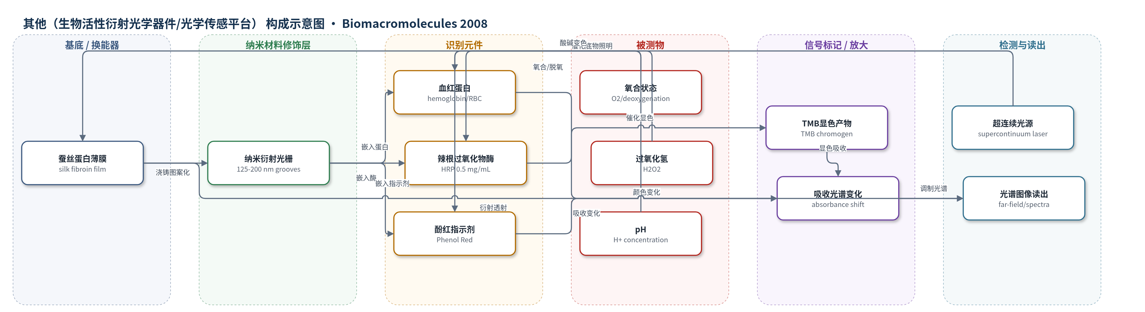
点击图片查看大图 · 依据论文自动绘制
传感器类型
其他(生物活性衍射光学器件/光学传感平台)
检测对象
氧合/脱氧血红蛋白(hemoglobin, Hb;氧状态 O2)、过氧化氢(hydrogen peroxide, H2O2)、pH(氢离子浓度);样品基质:蒸馏水、人血/红细胞-蚕丝膜、TMB底物溶液、pH缓冲液(1 mM NaOH、1 mM HCl、去离子水)。
检测原理
蚕丝蛋白水溶液浇铸到衍射光栅母版上,经干燥和水退火形成含β-折叠结构的透明蚕丝衍射光栅;生物分子在成膜前掺入并保留活性。当目标物改变时,嵌入生物分子的状态或反应产物改变:血红蛋白在氧/氮气氛下发生氧合/脱氧转变,吸收峰随之变化;辣根过氧化物酶催化过氧化氢氧化四甲基联苯胺,生成蓝/黄氧化产物,使吸收增强;酚红随pH改变颜色。这些吸收/颜色变化调制透过或衍射光的光谱。白光或超连续光源经蚕丝光栅衍射后,远场光谱、图像或功率计记录衍射级次和光谱变化,从而实现生物功能的光学读出。信号放大主要依赖酶催化TMB显色和衍射光栅的光谱分辨,而非HCR、RCA或CRISPR-Cas等核酸放大策略。
检测灵敏度
原文未报告LOD、线性范围、灵敏度斜率或相关系数。
效应效果
系统展示良好光学与生物功能稳定性:蚕丝膜可见光透射率90–95%,折射率约1.55,一级衍射效率34%(633 nm),表面粗糙度<10 nm rms,沟槽宽125–200 nm,可制备50×50 mm大面积光栅。血红蛋白掺杂膜4天后仍保留氧合/脱氧光谱响应,氮气脱氧15 min后氧合峰消失,恢复氧气后重现;HRP掺杂膜室温保存45天仍具TMB催化活性;酚红掺杂膜在不同pH下可逆改变衍射光谱,可重复使用。未报告选择性、抗干扰、RSD、回收率或与ELISA/HPLC/qPCR对比。作者强调其生物相容、可降解、力学强韧(蚕丝超薄膜弹性模量6.5 GPa、拉伸强度100 MPa),适合一次性/可降解生物传感器、植入和环境传感平台。
传感器的构成
- 基底/换能器:蚕丝蛋白薄膜(silk fibroin film),作为光学透明基质和衍射元件主体,折射率约1.55,厚度10–100 μm。
- 结构修饰:水退火诱导β-sheet结晶(β-sheet crystallinity),提高力学稳定性、不溶性和光学透明性。
- 纳米图案层:表面纳米沟槽/衍射光栅(groove width 125–200 nm,roughness <10 nm rms),实现白光衍射和光谱分辨。
- 生物识别/功能元件:血红蛋白/红细胞(hemoglobin/RBC),嵌入膜中保留氧合/脱氧吸收功能。
- 酶功能元件:辣根过氧化物酶(HRP),浓度0.5 mg/mL,催化TMB氧化显色。
- 小分子指示元件:酚红(Phenol Red/phenolsulfonphthalein),1 mg/mL,pH响应变色。
- 信号底物/标记:四甲基联苯胺(TMB),与H2O2在HRP催化下生成蓝/黄氧化产物,提供颜色/吸收信号。
- 光学读出:超连续白光激光/白光光源、吸收光谱/远场成像/热释电功率计,用于记录衍射光谱和衍射效率。
中文摘要
蚕丝基生物材料系统此前已被探索用于多种医疗和非医疗材料需求。蚕丝独特的生物物理特性使其能够生成高度定制的结构和形貌。为利用这些特性,作者优化了蚕丝蛋白的全水相处理,制备出新型表面纳米图案化蛋白质材料。通过控制纳米形貌,作者优化了蚕丝蛋白系统的光学特性,展示了表面形貌可控制至125 nm,并在大尺度上保持保真。表面纳米图案化使蚕丝蛋白可形成衍射光学元件,如衍射光栅、图案发生器和透镜,并通过控制β-折叠结晶度实现光学透明材料。此外,在形成蚕丝衍射光栅过程中掺入血红蛋白和过氧化物酶等生物组分。蚕丝蛋白在水中的环境处理与这些生物活性组分相结合,使嵌入分子保留活性,并为光学活性蚕丝膜提供附加功能和选择性。因此,生化与光学读出相结合是可行的,可在单一一次性/全可降解元件中同时实现光谱分辨和生物功能。这些新的表面纳米图案化、生物活性蚕丝蛋白基材料系统提供了独特组合,可能用于多种生物传感器需求,尤其结合其优异的力学性能、生物相容性和可控生物降解。
英文摘要
Silk-based biomaterial systems have been previously explored for a variety of medical and nonmedical materials needs. The unique biophysical features of silks provide options to generate highly tailored structures and morphologies with this unique family of fibrous proteins. To exploit these features, we have optimized the all aqueous processing of silk fibroin into novel surface nanopatterned protein materials. We have exploited control of this nanomorphology to optimize the optical features of these silk protein systems. We demonstrate control of surface morphology down to 125 nm, with fidelity over large length scales. This surface nanopatterning allows the silk protein to be formed into diffractive optics such as diffraction gratings, pattern generators, and lenses due to novel aqueous processing into optically clear materials via control of beta sheet crystallinity. Further, we incorporate biological components, such as hemoglobin and the enzyme peroxidase, during the process of forming the silk diffraction gratings. The ambient processing of the silk protein in water, in combination with these bioactive components, allows these entrained molecules to retain activity and provide added functions and selectivity to the optically active silk films. Thus, combinations of biochemical and optical readout is feasible and provides in a single, disposable/all degradable element with both spectral discrimination and biological function. These new surface nanopatterned, bioactive silk protein-based material systems offer a unique combination of features potentially useful for a range of biosensor needs, particularly when considered in concert with the remarkable mechanical properties of these proteins, their biocompatibility, and controllable biodegradation.