组成图示
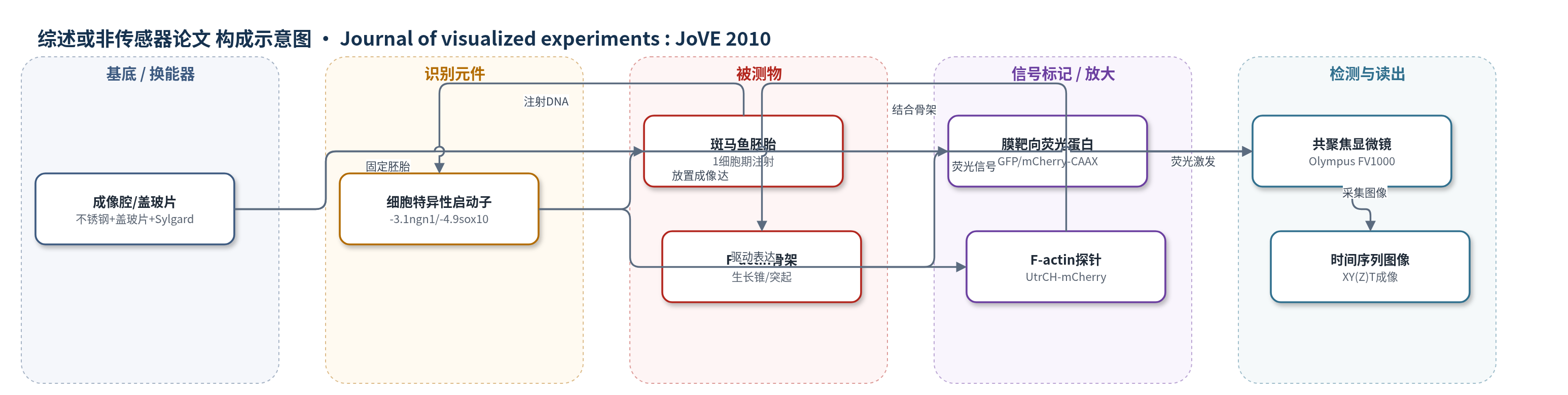
点击图片查看大图 · 依据论文自动绘制
传感器类型
综述或非传感器论文
检测对象
单个神经元(neurons)、神经嵴细胞(neural crest cells)的细胞运动与F-actin细胞骨架(F-actin cytoskeleton);样品基质:斑马鱼胚胎活体(zebrafish embryos)
检测原理
将含细胞特异性启动子(-3.1ngn1或-4.9sox10)的质粒DNA注射入1细胞期斑马鱼胚胎,形成镶嵌转基因表达。启动子仅在感觉神经元或神经嵴细胞中激活,驱动膜靶向GFP/mCherry或UtrCH-mCherry表达。UtrCH结构域结合F-actin,使mCherry荧光定位于F-actin富集区域,如生长锥和突起。共聚焦显微镜以低功率激光激发荧光,采集XY(Z)T时间序列图像。F-actin组装、解聚和分布变化引起荧光强度与空间分布改变,从而在活体中反映细胞运动、突起活动和轴突引导过程。
检测灵敏度
原文未报告 LOD、线性范围、灵敏度斜率或相关系数。
效应效果
方法在活体斑马鱼胚胎中实现单细胞标记,避免转基因整群标记;1细胞期注射0.5–1 nl、10–50 μg/ml质粒DNA,典型获得5–10%适合成像胚胎;过高表达导致毒性,需排除过亮胚胎;可在Tg(ngn1:gfp-caax)背景中注射25 pg ngn1:mCherry-caax单细胞红色标记;注射约20 pg ngn1:mCherry-UtrCH可在16.5–20 hpf观察生长锥F-actin;成像使用60X物镜(N.A.≥1.2),激光功率≤25%降低光损伤;作者主张用于神经嵴迁移、轴突引导和细胞运动机制研究。
传感器的构成
- 成像基底:不锈钢矩形、盖玻片、Sylgard硅橡胶、塑料环与真空脂,用于固定斑马鱼胚胎并适配倒置显微镜
- 样品介质:E3胚胎培养基、10 mM HEPES、0.02% Tricaine麻醉剂、低熔点琼脂糖,用于维持胚胎存活并固定成像
- 识别元件:细胞特异性启动子-3.1ngn1或-4.9sox10,驱动感觉神经元或神经嵴细胞中目的基因表达
- 信号标记物:膜靶向GFP/mCherry-CAAX或UtrCH-mCherry F-actin生物传感器探针,用于标记细胞膜或F-actin
- 换能读出:Olympus FV1000共聚焦显微镜、IX81显微镜、60X油镜/水镜,激光激发荧光并采集XY(Z)T时间序列
中文摘要
斑马鱼是活体观察发育过程中细胞行为的理想模型。其体外受精使胚胎各发育阶段均易于操作,光学透明性则允许在完整胚胎自然环境中高分辨率成像细胞与分子动态。本文采用活体成像方法分析神经嵴细胞迁移以及神经元轴突生长和引导过程中的细胞行为。活体成像有助于理解调控细胞运动机制;为观察突起活动和分子动态等细节,标记单个细胞具有优势。在斑马鱼中,质粒DNA注射可产生短暂镶嵌表达,相比其他细胞标记方法具有明显优势。转基因系常标记整个细胞群,可能掩盖单个细胞精细突起或分子分布变化;而1细胞期DNA注射比后期染料注射侵入性更小、更精确。本文描述标记发育中单个神经元或神经嵴细胞并在活体中成像其行为的方法:将质粒DNA注射入1细胞期胚胎,形成镶嵌转基因表达;载体含细胞特异性启动子,可在部分感觉神经元或神经嵴细胞中驱动目的基因表达。文中给出膜靶向GFP标记细胞,以及使用可在活细胞中可视化F-actin的生物传感器探针的示例。
英文摘要
The zebrafish is an ideal model for imaging cell behaviors during development in vivo. Zebrafish embryos are externally fertilized and thus easily accessible at all stages of development. Moreover, their optical clarity allows high resolution imaging of cell and molecular dynamics in the natural environment of the intact embryo. We are using a live imaging approach to analyze cell behaviors during neural crest cell migration and the outgrowth and guidance of neuronal axons. Live imaging is particularly useful for understanding mechanisms that regulate cell motility processes. To visualize details of cell motility, such as protrusive activity and molecular dynamics, it is advantageous to label individual cells. In zebrafish, plasmid DNA injection yields a transient mosaic expression pattern and offers distinct benefits over other cell labeling methods. For example, transgenic lines often label entire cell populations and thus may obscure visualization of the fine protrusions (or changes in molecular distribution) in a single cell. In addition, injection of DNA at the one-cell stage is less invasive and more precise than dye injections at later stages. Here we describe a method for labeling individual developing neurons or neural crest cells and imaging their behavior in vivo. We inject plasmid DNA into 1-cell stage embryos, which results in mosaic transgene expression. The vectors contain cell-specific promoters that drive expression of a gene of interest in a subset of sensory neurons or neural crest cells. We provide examples of cells labeled with membrane targeted GFP or with a biosensor probe that allows visualization of F-actin in living cells.