组成图示
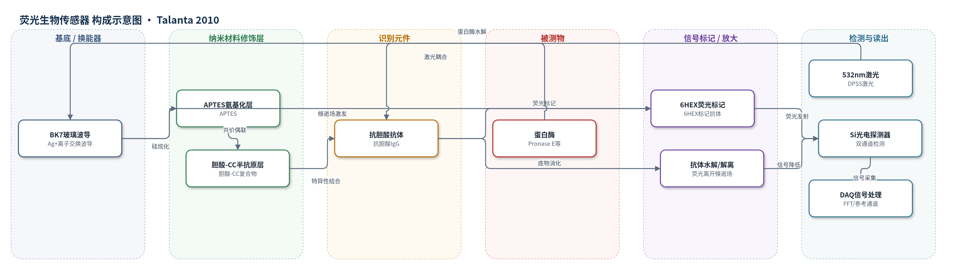
点击图片查看大图 · 依据论文自动绘制
传感器类型
荧光生物传感器
检测对象
蛋白酶(protease,如 Pronase E);样品基质:assay buffer(20 mM Tris-HCl、100 mM NaCl、5 mM CaCl2、50 nM ZnCl2,pH 7.3)
检测原理
APTES 氨基化 BK7 玻璃波导表面后,胆酸-半胱氨酸-三聚氯氰(CC)复合物经二氯三嗪与表面氨基共价结合,形成固定胆酸半抗原层。6HEX 标记抗胆酸抗体特异性结合半抗原,构成固相荧光蛋白酶底物。532 nm 激光经棱镜耦合进入波导,倏逝场激发近表面 6HEX 荧光,Si 光电探测器双通道记录。加入蛋白酶后,蛋白酶水解抗体多肽链,破坏抗体-半抗原结合,使荧光抗体从表面解离并离开倏逝场,荧光强度随时间下降。下降速率 Alfa 与蛋白酶活性在 0.03–2 units/mL 范围内线性相关;参考通道比值用于校正激光漂移和荧光漂白。
检测灵敏度
LOD: 0.0023 units/mL;线性范围: 0.03–2 units/mL;灵敏度斜率: 0.11 min−1/(units/mL);估计上限: 4 units/mL
效应效果
缓冲液对照未出现显著荧光下降,表明半抗原-抗体复合物在无蛋白酶时稳定。芯片经 pepsin 再生后连续 3 个循环信号一致,无背景累积,可重复使用。连续 16 h 激光脉冲下荧光漂白至 78%(通道1)和 82%(通道2),衰减率约 0.69%/h 和 0.56%/h;参考通道比值斜率为 3.6×10⁻⁵ min−1,与系统漂移 ±1×10⁻⁵ min−1 相当。系统噪声 0.001 V,信号 0.2 V。高浓度蛋白酶(≥0.5 units/mL)测量约 40 min,低浓度最长约 100 min。作者认为该系统可微型化、在线监测,适用于临床诊断、生物技术和食品/洗涤剂工业。
传感器的构成
- 基底/换能器:BK7 玻璃片经 Ag+ 离子交换制备 532 nm 单模平面波导,导光并形成倏逝场。
- 表面氨基化层:3-aminopropyl-triethoxysilane (APTES) 硅烷化玻璃表面,提供氨基用于共价偶联。
- 半抗原共价修饰层:cholic acid–cysteine–cyanuric chloride (CC) 复合物,经二氯三嗪与 APTES 氨基共价连接,固定胆酸半抗原。
- 识别/底物元件:6HEX 标记抗胆酸抗体 (anti-cholic acid IgG–6HEX),特异性结合胆酸半抗原并作为蛋白酶底物。
- 荧光标记物:6-carboxy-2’,4,4’,5’,7,7’-hexachlorofluorescein (6HEX) 共价标记抗体,提供倏逝场荧光信号。
- 样品反应层:蛋白酶样品(如 Pronase E)在 assay buffer 中接触芯片,水解抗体并降低荧光。
- 再生试剂:pepsin 消化剩余抗体,使芯片可重复加载。
- 读出系统:532 nm DPSS 激光、棱镜耦合、长通滤光片、Si 光电探测器与 DAQ,双通道记录荧光衰减。
中文摘要
荧光抗体蛋白(IgG)通过特异性结合共价固定在集成光学玻璃波导芯片表面的半抗原,被用作蛋白酶活性的测量底物。将芯片暴露于蛋白酶时,结合的荧光抗体分子被消化,倏逝场中可检测荧光按比例下降。该结合荧光抗体蛋白作为独特的通用蛋白酶底物,其生物活性与荧光信号共同构成测量基础。蛋白酶作用以实时模式监测,记录倏逝荧光的逐渐下降。芯片可通过过量胃蛋白酶完全消化抗体底物进行再生,并用新鲜标记抗体重新装载。该生物传感器用于检测多种蛋白酶活性,包括细菌蛋白酶制剂Pronase E。Pronase E活性的线性测量范围为0.03至2 units/mL。高蛋白酶浓度(≥0.5 units/mL)样品测量周期为40 min,较低浓度时测量时间最长可达100 min。该方法展示了实时蛋白酶生物传感的新原理。模块化集成光学玻璃波导生物传感器系统紧凑,由笔记本电脑控制,易于微型化并用作真正的探头器件,可应用于研究、临床诊断、生物技术加工以及食品和洗涤剂制造等领域。
英文摘要
Fluorescent antibody protein (IgG) was attached to the surface of an integrated optical glass waveguide chip via specific binding to a covalently attached hapten and used as a substrate for the measurement of protease activities. Exposure of the optical chip to proteases resulted in digestion of the bound fluorescent antibody molecules and proportional decrease in the detectable fluorescence resulting from loss of fluorescence from the evanescent field. The bound fluorescent antibody protein was used as a unique universal protease substrate in which the combined biological activity and fluorescence signal were the basis of measurement. The action of proteases was monitored in real-time mode where the gradual decrease in evanescent fluorescence was recorded. The chip was regenerated by complete digestion of the antibody substrate by excess pepsin and recharged by incubation with a fresh sample of the labelled antibody. The biosensor was used to detect activity of several proteases including a bacterial protease preparation, Pronase E. The linear range of measurable Pronase E activity was from 0.03 to 2 units/mL. A measurement cycle took 40 min for samples with high protease concentration (>or=0.5 units/mL), when the concentration of the protease was less measurement times up to 100 min were required. The method demonstrates the principle of a new mode of real-time biosensing of proteases. The modular integrated optical glass waveguide biosensor system used in this study is compact and controlled by a laptop computer and could easily be miniaturised and utilized as a true probe device for detecting proteases with potential applications in a wide range of areas including research, clinical diagnostics, biotechnology processing and food and detergent manufacturing industries.