组成图示
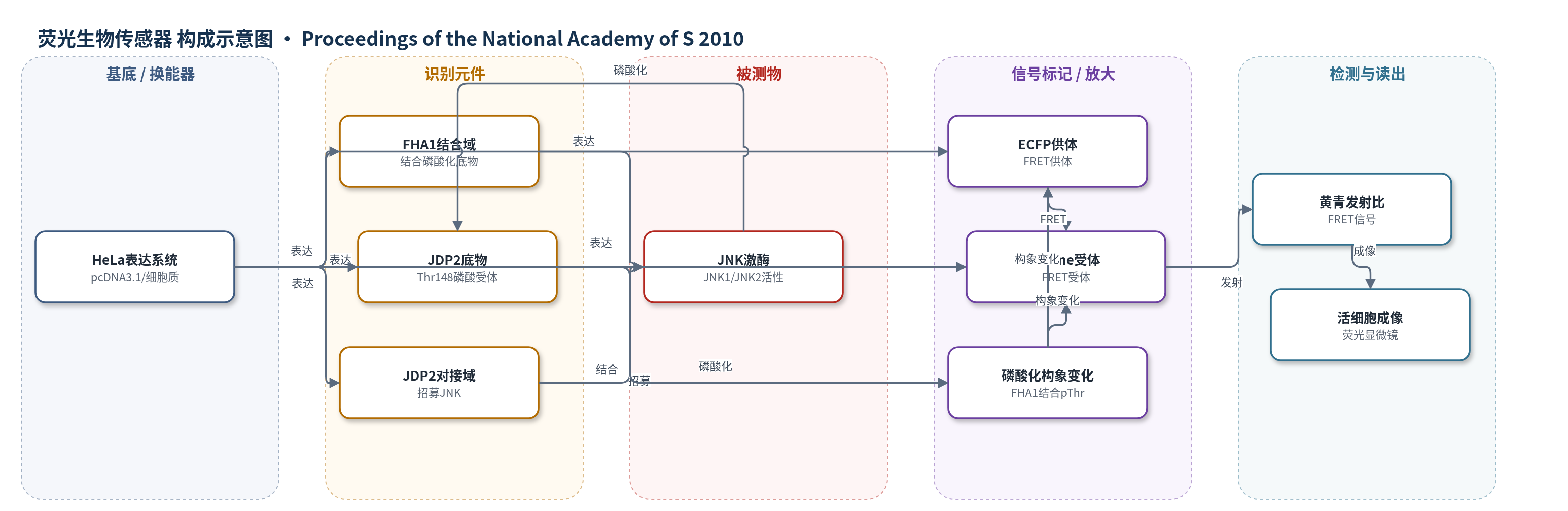
点击图片查看大图 · 依据论文自动绘制
传感器类型
荧光生物传感器
检测对象
JNK 活性(JNK activity);样品基质:HeLa 活细胞(胞质、细胞核、线粒体、质膜)
检测原理
JNKAR1 由 ECFP、FHA1、JDP2 底物、JDP2 对接域和 citrine 串联组成。JNK 通过对接域被招募到传感器附近,并特异性磷酸化 JDP2 底物中的 Thr-148。磷酸化后,FHA1 与磷酸化底物结合,使 ECFP 与 citrine 的相对距离和取向改变,FRET 效率升高,表现为黄/青发射比增加。该比值随 JNK 活性增强而升高,在 anisomycin、sorbitol 或 TNF-α 刺激下可升高 15%–30%。荧光显微镜通过双发射比成像连续读出信号,从而在活细胞中定量 JNK 活性的时空动态。
检测灵敏度
原文未报告 LOD、线性范围、灵敏度斜率或相关系数。
效应效果
JNKAR1 对 JNK 具有选择性:MEK 抑制剂 U0126 和 p38 抑制剂 SB203580 不改变 anisomycin 响应,而 JNK 抑制剂 VIII 消除响应;PDGF 激活 ERK 时不响应。5 μM anisomycin 使黄/青比升高 20.0±6.6%,t1/2 为 18.7±3.6 min;500 nM 升高 19.8±8.7%。核、线粒体和质膜靶向版本分别升高 30.8±5.0%、11.0±3.5% 和 13.4±0.3%。信号可逆,60–90 min 恢复基线并可再次响应。单细胞分析显示 100 nM anisomycin 产生双峰响应(P<0.001),Hill 系数达 9,与 Imstain 检测的 phospho-ATF-2 双峰(Hill 17.2)一致,支持 JNK 双稳态。
传感器的构成
- 表达载体:pcDNA 3.1 质粒,携带 JNKAR1 编码序列并在 HeLa 细胞中表达
- 供体荧光蛋白:ECFP,作为 FRET 供体,提供青色荧光
- 受体荧光蛋白:citrine,作为 FRET 受体,提供黄色荧光
- 磷酸化结合域:FHA1,识别并结合磷酸化 JDP2 底物,引起构象变化
- 底物识别元件:JDP2 基序(Thr-148 磷酸受体),被 JNK 磷酸化
- 对接域:JDP2 docking domain,招募 JNK 提高磷酸化特异性与效率
- 连接肽:AKAR2 同源 linker(SAGKPGSGEGSTKGLV),连接 FHA1 与底物
- 定位序列:NLS、DAKAP1a 或 Lyn 片段,将传感器靶向细胞核、线粒体或质膜
中文摘要
JNK 介导的信号通路可将应激刺激和细胞因子等信号转导为凋亡、增殖、分化与炎症等功能反应。为在细胞内环境中刻画该通路的动态信息流与信号处理,作者构建了一种基因编码的荧光蛋白生物传感器 JNKAR1(JNK activity reporter),用于检测内源性 JNK 活性。该传感器基于磷酸化依赖的 FRET 变化:当 JNK 磷酸化其底物序列后,磷酸化氨基酸结合域 FHA1 与磷酸化底物结合,改变青色荧光蛋白 ECFP 与黄色荧光蛋白 citrine 之间的距离和取向,使黄/青发射比升高 15%–30%。JNKAR1 可检测核糖体毒性应激、渗透压应激及 TNF-α 诱导的 JNK 活性,并在胞质、细胞核、线粒体和质膜中显示相似动力学。单细胞定量分析表明,anisomycin 诱导的 JNK 活性具有超敏感性、持续性和双峰性,符合双稳态系统特征。该传感器为在单个活细胞中评估 JNK 级联的信号特性奠定了基础。
英文摘要
The signaling pathway mediated by JNK transduces different types of signals, such as stress stimuli and cytokines, into functional responses that mediate apoptosis, as well as proliferation, differentiation, and inflammation. To better characterize the dynamic information flow and signal processing of this pathway in the cellular context, a genetically encoded, fluorescent protein-based biosensor was engineered to detect endogenous JNK activity. This biosensor, named JNKAR1 (for JNK activity reporter), specifically detects stress- (ribotoxic and osmotic) and cytokine- (TNF-alpha) induced JNK activity in living cells with a 15 to 30% increase in the yellow-to-cyan emission ratio because of a phosphorylation-dependent increase in FRET between two fluorescent proteins. JNK activity was detected not only in the cytoplasm, but also in the nucleus, mitochondria, and plasma membrane with similar kinetics after induction of ribotoxic stress by anisomycin, suggesting relatively rapid signal propagation to the nuclear, mitochondrial, and plasma membrane compartments. Furthermore, quantitative single-cell analysis revealed that anisomycin-induced JNK activity exhibited ultrasensitivity, sustainability, and bimodality, features that are consistent with behaviors of bistable systems. The development of JNKAR1, therefore, laid a foundation for evaluating the signaling properties and behaviors of the JNK cascade in single living cells.