组成图示
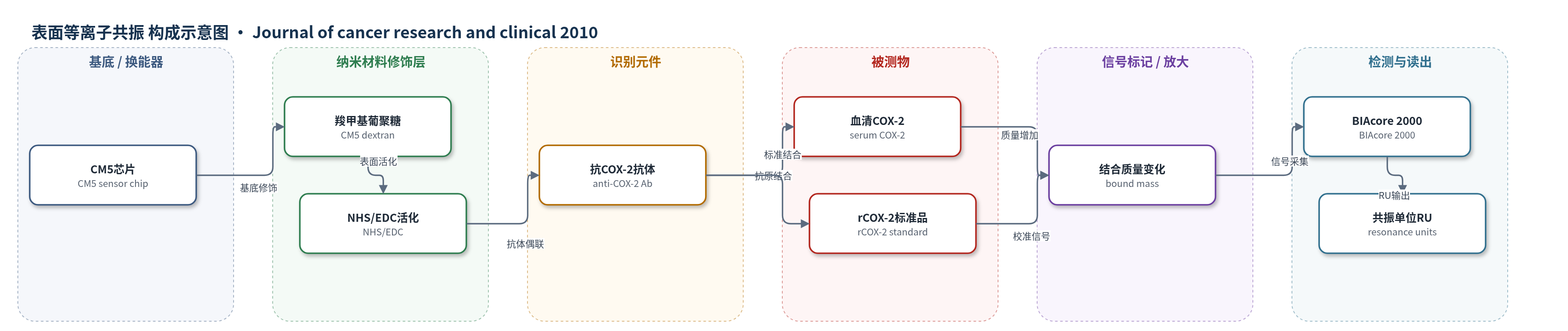
点击图片查看大图 · 依据论文自动绘制
传感器类型
表面等离子共振(SPR)生物传感器
检测对象
血清 COX-2(cyclooxygenase-2, COX-2),样品基质:人血清(serum)
检测原理
该SPR生物传感器以CM5芯片为基底,其表面羧甲基葡聚糖经NHS/EDC活化后,通过胺基共价偶联固定anti-COX-2抗体,并用乙醇胺封闭残余活性位点。检测时,血清样品中的COX-2与芯片表面抗体特异性结合,结合蛋白质量改变芯片表面附近介质的折射率,使表面等离子体共振角发生偏移。BIAcore 2000将共振角变化转换为共振单位(RU),RU值与结合质量及COX-2浓度成正比。实验以rCOX-2建立标准曲线,将血清稀释1:99后流过芯片,由RU值反推血清COX-2浓度。该法无需标记或酶放大,属于直接质量传感;每次结合后用1 mM NaOH和10 mM glycine-HCl再生表面。
检测灵敏度
标准曲线浓度范围: 4.4–44.0 μg(rCOX-2);血清稀释1:99,RU范围: 19,451.7–20,090.8(原文称处于标准曲线线性范围内)
效应效果
SPR法检测76例口腔癌与43例正常人血清COX-2,患者组9.4±4.4 μg/ml,高于正常组3.5±1.3 μg/ml(P<0.0001,约3倍);晚期组10.5±4.4 μg/ml高于早期组5.8±1.6 μg/ml(P<0.0004),淋巴结转移组10.4±4.9 μg/ml高于无转移组7.2±1.8 μg/ml(P<0.002)。血清水平与组织免疫组化一致。肽抑制剂P1/P4/P6诱导KB细胞凋亡最高49.6%(对照2.0%),对MCF-7无作用,作者认为SPR具临床应用潜力。
传感器的构成
- 基底/换能器:CM5传感器芯片(CM5 sensor chip),表面为羧甲基葡聚糖,承载SPR信号
- 化学活化层:NHS与EDC活化羧甲基葡聚糖表面,形成NHS酯用于抗体胺基偶联
- 识别元件:anti-COX-2抗体(anti-COX-2 antibody),固定于芯片表面特异性捕获COX-2
- 封闭剂:ethanolamine(乙醇胺),灭活残余NHS酯,减少非特异性结合
- 信号读出:BIAcore 2000 SPR仪器,通过共振角变化记录共振单位(RU)
中文摘要
本研究旨在定量检测烟草相关口腔癌患者循环中的COX-2水平,并评估COX-2肽抑制剂对KB口腔癌细胞的体外抗肿瘤活性。作者采用新型生物传感器表面等离子共振(SPR)技术,检测76例口腔癌患者和43例正常对照的血清COX-2水平;同时用碘化丙啶(PI)标记流式细胞术、Alamar Blue、MTS和Annexin V结合实验评价5种COX-2抑制肽的抗肿瘤作用。结果显示,口腔癌患者血清COX-2水平较正常对照升高约3倍(P<0.0001);晚期肿瘤和淋巴结转移患者血清COX-2水平显著升高。循环COX-2水平较高的患者,其病变组织抗COX-2抗体免疫反应性也较高。肽抑制剂显著降低肿瘤细胞活力,抑制生长和增殖,并诱导细胞毒性和凋亡;但对正常人类白细胞及不表达COX-2的MCF-7细胞无类似作用。结论认为,SPR可作为定量评估COX-2的蛋白质组学技术,用于识别高危口腔癌前病变或隐匿癌患者,并监测COX-2靶向治疗反应;COX-2肽抑制剂可能是人口腔癌的新型强效抗肿瘤药物。
英文摘要
PURPOSE: The aim of this study was to quantitate circulating COX-2 levels in patients with tobacco-related intraoral cancer and to evaluate antitumor activities of COX-2 peptide inhibitors in vitro on KB cell lines.
PATIENTS AND METHODS: We used a novel biosensor-based surface plasmon resonance (SPR) technique for estimation of circulating COX-2 levels in 76 patients with oral cancer and 43 normal individuals. Antitumor activities of five COX-2 inhibitory peptides were evaluated using propidium iodide labeling and flow cytometry, alamar blue, MTS, and annexin-V binding assays.
RESULTS: Patients with oral cancer showed threefold increase in serum COX-2 level when compared to normal controls (P < 0.0001). Further, late-stage tumors and lymph node metastasis were associated with significant increase in serum COX-2 levels. Patients with higher circulating COX-2 also showed higher immunoreactivity to anti-COX-2 antibody in the lesions. The peptides significantly reduced viability and inhibited growth/proliferation, induced cytotoxicity and apoptosis in tumor cells. However, no such effect was observed either on normal human leukocytes or on MCF-7 cell line that did not over express COX-2.
CONCLUSION: Our results indicate that SPR may be a useful proteomic technique for quantitative assessment of COX-2 and to identify patients with high-risk oral premalignant or occult cancer, as well as in monitoring response to novel COX-2 targeting strategies. Furthermore, COX-2 peptide inhibitors appear to be a new class of potent anticancer agent for human oral carcinoma.