组成图示
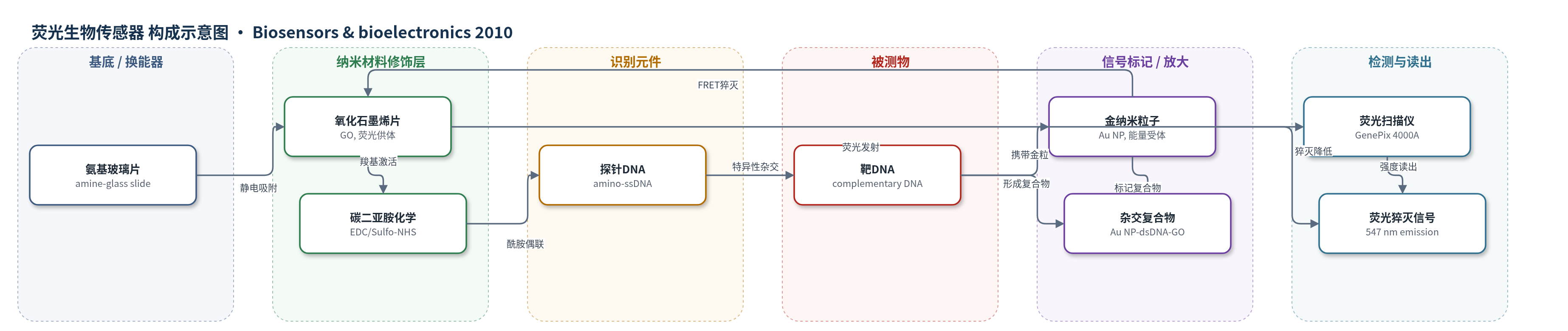
点击图片查看大图 · 依据论文自动绘制
传感器类型
荧光生物传感器
检测对象
特定互补 DNA(complementary target DNA, Au NP-labeled target DNA);样品基质:DNA 寡核苷酸溶液/微阵列点样(0.1 M MES 缓冲液、去离子水)
检测原理
GO 因氧化引入的 sp3 缺陷域中电子-空穴复合而产生约 547 nm 荧光。探针 DNA 通过 EDC/Sulfo-NHS 共价固定于 GO 表面,作为识别元件。Au NP 标记的互补靶 DNA 与探针杂交后,Au NP 被拉近至 GO 表面,形成 GO-Au NP 供体-受体对。由于 Au NP 具有强非辐射能量接受能力,GO 激发能量通过 Förster 共振能量转移(FRET)转移至 Au NP 并耗散,导致 GO 荧光猝灭。靶 DNA 浓度越高,杂交形成的 Au NP 数量越多,猝灭效率越高,荧光信号越低。最终由荧光扫描仪读取阵列点荧光强度变化,实现特定 DNA 杂交的定量识别。
检测灵敏度
LOD: ~0.1 pmol (200 nM);线性范围: 4 pmol (8 μM)–10 amol (20 nM)
效应效果
该传感器在四重复实验中表现出浓度依赖的荧光猝灭:4、2、1、0.1、0.01 pmol 靶 DNA 对应猝灭效率 87±6%、74±8%、67±11%、47±2%、32±4%。非互补 DNA 对照与原始 GO 荧光差异小于 8.3%,说明杂交特异性较好。作者以 50% 猝灭为阈值估算 LOD 约 0.1 pmol(200 nM),认为与传统 DNA 微阵列常用量相当。溶液相中 Au NP 标记双链 DNA 使 GO 荧光降低超过 95%;HRTEM 显示 Au NP 密度 70–120 个/μm²,平均约 80 个/μm²。作者主张该 GO 荧光阵列可拓展至纳米医学、纳米生物技术和免疫分析。
传感器的构成
- 基底/换能器:氨基修饰玻璃片(amine-modified glass slide),带正电荷,静电吸附 GO 片并形成阵列,支撑传感界面。
- 纳米材料修饰层:氧化石墨烯片(graphene oxide, GO),提供约 547 nm 荧光发射和表面羧基连接位点。
- 连接层:EDC 与 Sulfo-NHS 碳二亚胺化学,激活 GO 羧基形成 NHS 酯,偶联氨基修饰 DNA。
- 识别元件:氨基修饰单链探针 DNA(amino-modified ssDNA, 5′-H2N-TTT TTT GCA CAC GCG CAC-3′),共价固定于 GO,特异性识别互补靶 DNA。
- 信号标记物:金纳米粒子标记靶 DNA(Au NP-labeled target DNA, 5′-SH TTT TTT GTG CGC GTG TGC-3′),杂交后 Au NP 作为 FRET 能量受体猝灭 GO 荧光。
- 杂交/洗涤介质:0.1 M MES 缓冲液和去离子水(DI water),用于点样、杂交和去除非特异结合。
- 读出层:Axon GenePix 4000A 荧光扫描仪,读取 GO 阵列 547 nm 荧光强度变化。
中文摘要
氧化石墨烯(GO)具有水分散性、表面可修饰和光致发光等特性,适合用于生物传感。本研究将 GO 片以阵列形式固定于氨基玻璃片上,利用 EDC/Sulfo-NHS 碳二亚胺化学将氨基修饰探针 DNA 共价连接至 GO 表面羧基。当金纳米粒子(Au NP)标记的互补靶 DNA 与探针杂交时,Au NP 作为能量受体靠近 GO 荧光供体,通过 Förster 共振能量转移(FRET)猝灭 GO 约 547 nm 的荧光发射。HRTEM 显示杂交后 Au NP 主要分布于 GO 边缘和褶皱结构,平均密度约 80 个/μm²,最高猝灭效率约 87%。该 GO 阵列可依据荧光强度变化识别特定 DNA 杂交,为 DNA 检测、分子成像和纳米生物技术提供低成本、可放大的传感平台。
英文摘要
The unique properties of graphene oxides (GO) such as water dispersibility, versatile surface modification, and photoluminescence make them suitable for biological applications. In this study, we explored the use of GO sheets as a novel DNA biosensor by applying the GO in an array format to recognize specific DNA-DNA hybridization interaction. When the probe DNA linked to the surface of GO by using carbodiimide chemistry is hybridized with a gold nanoparticle (Au NP) labeled complementary DNA strand, the fluorescence emission intensity of the GO array is drastically reduced. TEM data reveal that the Au NPs are dispersed on the GO surface, particularly at edges and folded structures upon hybridization with a density of approximately 80 Au NPs per microm(2). This leads to ca. 87% fluorescence quenching as a consequence of fluorescence energy transfer between Au NPs and the GO sheets. These results suggest that the GO nanomaterials, which are readily synthesized on a large scale from a cheap graphite source, could have a wide range of bioapplications in the fields of biosensors, molecular imaging and nanobiotechnology.