组成图示
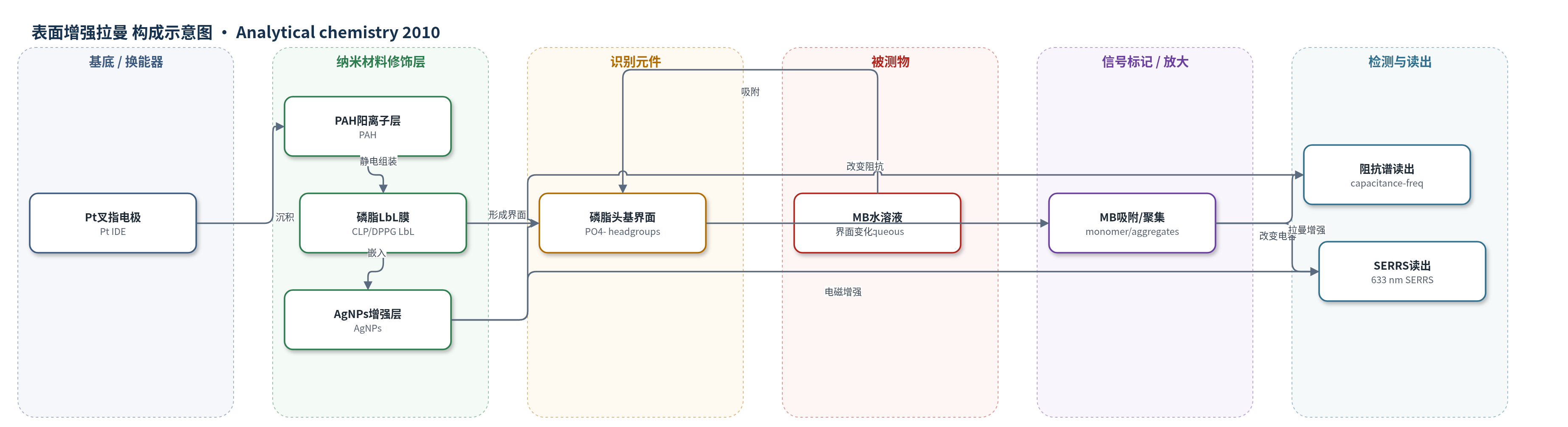
点击图片查看大图 · 依据论文自动绘制
传感器类型
表面增强拉曼(SERS)生物传感器
检测对象
亚甲基蓝(methylene blue, MB),水溶液(超纯水/MB 水溶液)
检测原理
MB 为阳离子吩噻嗪染料,进入水溶液后与磷脂 LbL 膜表面的阴离子头基(PO4-)发生静电吸附,形成膜/溶液界面识别事件。随着 MB 浓度升高,界面未平衡电荷、双电层结构和膜内电荷分布发生变化,使 Pt 叉指电极的电容-频率阻抗响应改变,从而通过交流阻抗谱读出浓度信息。同时,嵌入磷脂膜中的 AgNPs 在 633 nm 激光下产生局域表面等离子体增强,并与 MB 的紫外-可见吸收共振,形成 SERRS 信号;MB 吸附量及其单体/聚集体状态决定拉曼峰强度与峰位,实现痕量检测与结构分辨。
检测灵敏度
检测下限: 10^-11 M
效应效果
在 10^-7、10^-9 和 10^-11 M 的 MB 水溶液中,阻抗谱电容-频率响应随浓度变化,PCA 图可区分裸电极与四种 LbL 膜传感单元,并显示 MB 浓度增加时数据点呈清晰趋势;AgNPs 的引入对 PC1 贡献显著。SERRS 在 10^-11 M 下仍可检测 MB,并能区分 MB 单体与聚集体。作者估算 SERRS/RRS 增强因子约为 2×10^3,虽比先前工作低约一个数量级,但与电磁机制预测一致。该耦合方法兼具高灵敏度与分子结构信息,适用于生物膜模拟系统中痕量分子相互作用研究。
传感器的构成
- 基底/换能器电极:Pt 叉指电极(Pt interdigitated electrodes),承载 LbL 膜并用于交流阻抗谱与原位 SERRS 测量。
- 阳离子聚电解质层:PAH(poly(allylamine hydrochloride)),提供 NH3+ 位点,与阴离子磷脂静电组装形成 LbL 膜。
- 磷脂膜层:CLP(cardiolipin)或 DPPG(dipalmitoyl phosphatidyl glycerol),形成生物膜模拟 LbL 膜,作为传感/换能界面。
- 纳米增强层:AgNPs(silver nanoparticles),分散于磷脂 LbL 膜中,增强 MB 的 SERRS 信号并改变界面电容。
- 识别/吸附层:磷脂阴离子头基(PO4-)与 MB 阳离子静电吸附,实现 MB 在膜/溶液界面的捕获。
- 信号读出:阻抗分析仪(Solartron 1260A)读取电容-频率;微拉曼光谱仪(Renishaw inVia,633 nm)读取 SERRS 谱。
中文摘要
表面增强拉曼散射(SERS)及传感器/生物传感器分析常用于研究药物-生物分子相互作用或痕量分析物检测。本研究将表面增强共振拉曼散射(SERRS)与基于阻抗谱的电子舌系统相结合,以同时获得高灵敏度与结构层面信息。作者利用磷脂层状自组装(LbL)膜作为生物膜模拟系统,将心磷脂(CLP)和二棕榈酰磷脂酰甘油(DPPG)作为换能材料修饰在铂叉指电极上,构成传感单元阵列。该电子舌系统可在亚纳摩尔浓度下检测吩噻嗪染料亚甲基蓝(MB)。SERRS 用于研究 MB 与磷脂接触时的分子排列(单体或聚集体)。关键在于将银纳米颗粒(AgNPs)吸附在磷脂 LbL 膜内;该策略未损害电子舌性能,并可在 MB 水溶液浸泡后对 LbL 膜进行原位 SERRS 谱采集。通过 SERRS 检测 MB 的结果与文献报道相似,但具有前所未有的灵敏度。
英文摘要
The surface-enhanced Raman scattering (SERS) effect and sensor and biosensor analyses are widely applied to investigate drug-biomolecule interactions or to detect trace amount of analytes. In this work, surface-enhanced resonance Raman scattering (SERRS) and an electronic tongue system using impedance spectroscopy were brought together, combining sensitivity and structural level information. Taking advantage of the use of layer-by-layer (LbL) films of phospholipids as biological membrane mimetic systems, cardiolipin (CLP) and dipalmitoyl phosphatidyl glycerol (DPPG) were applied as transducers onto Pt interdigitated electrodes forming an array of sensing units. This e-tongue system was able to detect the phenothiazine methylene blue (MB) below nanomolar concentrations. SERRS was applied to investigate the MB molecular arrangement (monomers or aggregates) when in contact with the phospholipids at trace levels of concentration. The key point was the adsorption of Ag nanoparticles (AgNPs) within the phospholipid LbL films. This approach did not compromise the e-tongue performance and allowed the recording of in situ SERRS spectra for the LbL films after immersion into MB aqueous solutions. The detection of MB through SERRS gave similar results to those reported in the literature but now with an unprecedented sensitivity.