组成图示
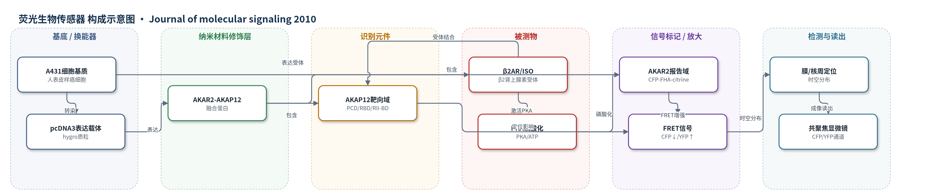
点击图片查看大图 · 依据论文自动绘制
传感器类型
荧光生物传感器
检测对象
AKAP12磷酸化(phosphorylation of AKAP12 by PKA);样品基质:人表皮样癌细胞A431活细胞
检测原理
β2肾上腺素受体(β2AR)被异丙肾上腺素激活后,经Gs-腺苷酸环化酶-cAMP通路激活PKA。AKAR2-AKAP12中AKAR2模块含有PKA底物序列、FHA磷酸氨基酸结合域、CFP和citrine YFP。PKA磷酸化AKAR2底物序列后,FHA域与磷酸化序列紧密结合,使CFP与citrine YFP距离缩短,产生FRET,表现为CFP荧光下降、YFP荧光上升;磷酸酶去磷酸化则FRET减弱。AKAP12模块通过PCD靶向膜、RBD结合β2AR、RII-BD锚定PKA,使报告域定位于支架所在微区。因此FRET强度随局部磷酸化AKAP12比例变化,并可反映膜区与核周胞质区的时空动态。
检测灵敏度
原文未报告LOD、线性范围、灵敏度斜率或R^2。
效应效果
AKAR2-AKAP12在A431细胞中表达后保留AKAP12功能,可挽救AKAP12敲低细胞中β2AR再敏化与回收缺陷。FRET信号具PKA依赖性:KT5720消除异丙肾上腺素诱导的FRET;AKAR2 T/P突变无FRET;HT-31肽阻断PKA RII-AKAP结合后FRET消失,HT-31p不阻断。β激动刺激使膜AKAP12增加3-5倍,膜区FRET 5 min内上升并稳态,核周胞质FRET 30 min内渐进增加。morpholino敲低内源AKAP12>85%(部分>90%)。未报告RSD和加标回收率,作者认为其可活细胞时空监测AKAP12磷酸化与GPCR再敏化/回收。
传感器的构成
- 融合蛋白主体:AKAR2-AKAP12,N端AKAR2与AKAP12融合,作为PKA磷酸化报告器
- AKAR2报告模块:CFP、PKA底物序列、FHA磷酸氨基酸结合域、citrine YFP,磷酸化后产生CFP-YFP FRET
- AKAP12靶向模块:PCD、RBD、RII-BD,锚定PKA、结合β2AR并靶向细胞膜
- 表达载体:pcDNA3 hygro质粒,携带AKAR2-AKAP12 cDNA,用于A431细胞表达
- 细胞基质:人表皮样癌细胞A431,提供β2AR、PKA和cAMP信号环境
- 刺激配体:异丙肾上腺素(isoproterenol),激活β2AR-PKA通路
- 读出系统:Zeiss LSM 510共聚焦显微镜,CFP 473-495 nm与YFP/citrine 527-591 nm检测FRET
中文摘要
蛋白激酶A(PKA)在几乎所有细胞信号转导中起核心作用,但其在活细胞中的时空激活动态难以直接观察。遗传编码荧光探针AKAR2可响应PKA磷酸化并产生荧光共振能量转移(FRET)信号,是PKA活性的生物传感器。AKAP12是一种支架蛋白,可锚定PKA、G蛋白偶联受体(GPCR)和膜磷脂,并催化受体再敏化与回收。本研究将AKAR2融合至AKAP12 N端,构建AKAR2-AKAP12融合蛋白,以评估其能否在保留AKAP12功能的同时报告该支架的动态磷酸化。结果显示,AKAR2-AKAP12可在哺乳动物细胞中表达并完全保留功能,在人表皮样癌细胞A431中响应β肾上腺素能刺激,揭示AKAP12被PKA磷酸化的时空激活过程。结论表明,该融合蛋白可“生物感知”AKAP12磷酸化:支架与细胞膜结合并快速被PKA磷酸化,核周胞质中磷酸化支架的积累反映PKA激活形式的AKAP12,其催化失敏内化GPCR的再敏化与回收。
英文摘要
BACKGROUND: The cAMP-dependent protein kinase A (PKA) plays a pivotal role in virtually all cells, there being a multitude of important target molecules that are substrates for PKA in cell signaling. The spatial-temporal dynamics of PKA activation in living cells has been made accessible by the development of clever biosensors that yield a FRET signal in response to the phosphorylation by PKA. AKAR2 is genetically encoded fluorescent probe that acts as a biosensor for PKA activation. AKAP12 is a scaffold that docks PKA, G-protein-coupled receptors, cell membrane negatively-charged phospholipids, and catalyzes receptor resensitization and recycling. In the current work, the AKAR2 biosensor was fused to the N-terminus of AKAP12 to evaluate its ability to function and report on dynamic phosphorylation of the AKAP12 scaffold.
RESULTS: AKAR2-AKAP12 can be expressed in mammalian cells, is fully functional, and reveals the spatial-temporal activation of AKAP12 undergoing phosphorylation by PKA in response to beta-adrenergic activation in human epidermoid carcinoma A431 cells.
CONCLUSION: The dynamic phosphorylation of AKAP12 "biosensed" by AKAR2-AKAP12 reveals the scaffold in association with the cell membrane, undergoing rapid phosphorylation by PKA. The perinuclear, cytoplasmic accumulation of phosphorylated scaffold reflects the phosphorylated, PKA-activated form of AKAP12, which catalyzes the resensitization and recycling of desensitized, internalized G-protein-coupled receptors.