组成图示
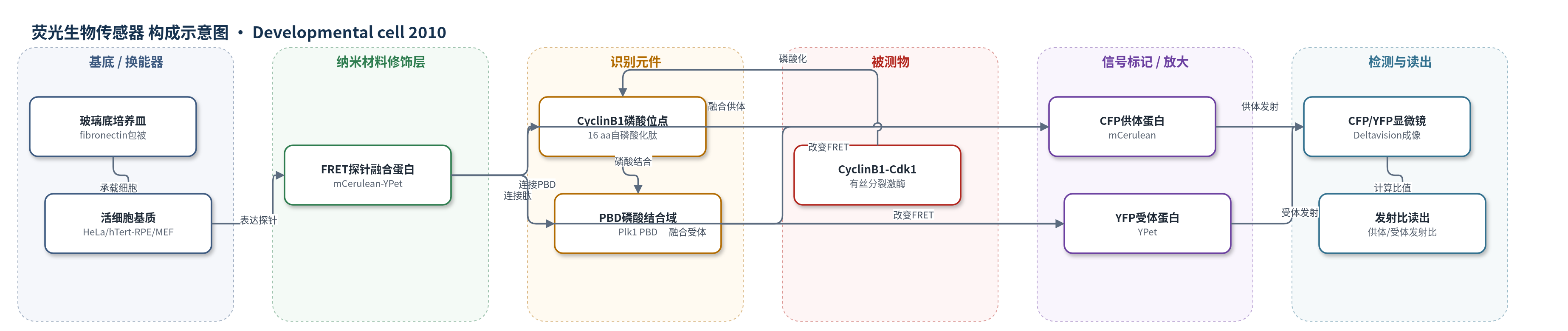
点击图片查看大图 · 依据论文自动绘制
传感器类型
荧光生物传感器
检测对象
CyclinB1-Cdk1激酶活性(CyclinB1-Cdk1 kinase activity);样品基质:活人细胞(HeLa、hTert-RPE、MEF)
检测原理
该传感器为基因编码FRET探针:mCerulean供体与YPet受体之间连接人CyclinB1自磷酸化位点肽段和Plk1 PBD。当CyclinB1-Cdk1激酶活性升高时,其特异性磷酸化连接肽;PBD随即结合磷酸化肽,使探针构象改变,缩短或优化供体-受体距离,提高FRET效率。荧光显微镜分别采集CFP供体发射和YFP受体发射,计算donor/acceptor emission ratio;比值升高代表激酶活性升高,比值下降代表磷酸酶去磷酸化或Cdk抑制。信号反映CyclinB1-Cdk1与拮抗磷酸酶之间的净平衡,而非单纯底物浓度,因此可实时监测活细胞中有丝分裂进入的激酶动力学。
检测灵敏度
原文未报告LOD、线性范围、灵敏度斜率或相关系数。
效应效果
该传感器选择性良好:G1、早S和G2期FRET不变,CyclinA-Cdk、Plk1、Aurora B、MAP激酶抑制剂不直接改变信号;Cdk抑制剂可立即逆转前期/中期信号。Plk1抑制剂延迟有丝分裂进入,但不改变CyclinB1-Cdk1激活动力学。HeLa中激活早于NEBD 27±7 min,RPE为18±6 min,活性约30 min升至峰值。低FRET增幅(<10%)可触发细胞圆化、核输入和CyclinB1降解,约15%增幅才触发核仁解体和NEBD。
传感器的构成
- 成像基底:玻璃底培养皿(glass-bottom dish,fibronectin包被),承载活细胞并支持时间序列荧光成像
- 表达基质:HeLa、hTert-RPE或MEF活细胞,电穿孔表达FRET探针并提供细胞内环境
- FRET换能器:mCerulean(CFP)与YPet(YFP)荧光蛋白对,作为供体/受体产生可比率荧光信号
- 磷酸化识别肽:人CyclinB1自磷酸化位点16 aa连接肽(-1位Ser突变),被CyclinB1-Cdk1特异性磷酸化
- 磷酸结合域:Plk1的polo box domain(PBD),结合磷酸化连接肽并诱导构象变化
中文摘要
CyclinB1-Cdk1激酶是有丝分裂促进因子(MPF)的核心催化活性,但其在哺乳动物细胞中何时以及以何种速度激活仍不清楚。作者开发了一种特异性识别CyclinB1-Cdk1的FRET生物传感器,可在活人细胞中以高时间分辨率检测其激酶活性。该传感器由mCerulean供体、YPet受体、CyclinB1自磷酸化位点连接肽和Plk1 polo box domain(PBD)组成;当CyclinB1-Cdk1磷酸化连接肽后,PBD结合磷酸肽并改变FRET效率,从而通过供体/受体发射比读出活性。结果显示,CyclinB1-Cdk1在G2期无活性,在核膜破裂(NEBD)前约27±7 min(HeLa)或18±6 min(RPE)开始激活,并在约30 min内逐步升至峰值。不同水平的激酶活性依次触发细胞圆化、核输入、APC/C激活、核仁解体和NEBD,揭示了有丝分裂进入时细胞重排的协调机制。
英文摘要
The CyclinB1-Cdk1 kinase is the catalytic activity at the heart of mitosis-promoting factor (MPF), yet fundamental questions concerning its role in mitosis remained unresolved. It is not known when and how rapidly CyclinB1-Cdk1 is activated in mammalian cells, nor how its activation coordinates the substantial changes in the cell at mitosis. Here, we have developed a FRET biosensor specific for CyclinB1-Cdk1 that enables us to assay its activity with very high temporal precision in living human cells. We show that CyclinB1-Cdk1 is inactive in G2 phase and activated at a set time before nuclear envelope breakdown, thereby initiating the events of prophase. CyclinB1-Cdk1 levels rise to their maximum extent over the course of approximately 30 min, and we demonstrate that different levels of CyclinB1-Cdk1 kinase activity trigger different mitotic events, thus revealing how the remarkable reorganization of the cell is coordinated at mitotic entry.