组成图示
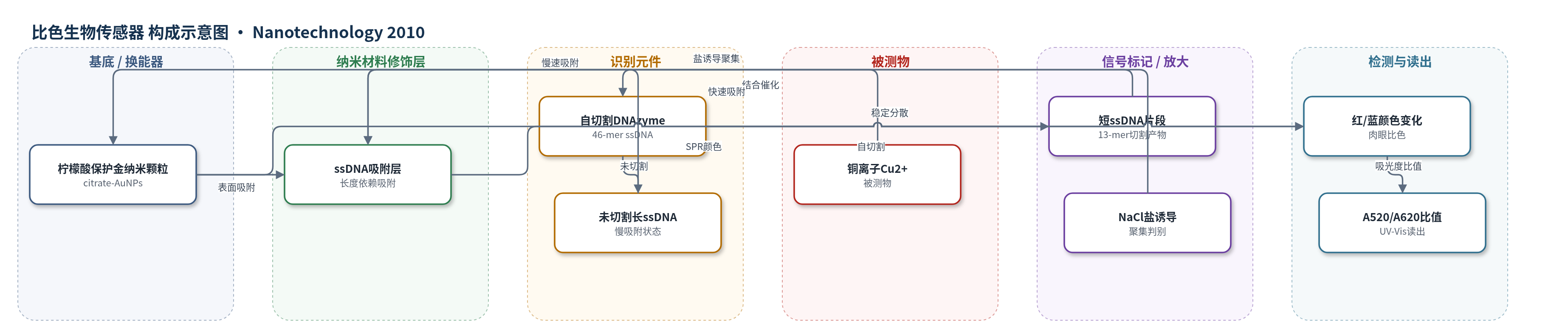
点击图片查看大图 · 依据论文自动绘制
传感器类型
比色生物传感器
检测对象
铜(II)离子(Cu2+),样品基质为湖水/水样(加标水样)
检测原理
该传感器以单分子Cu2+依赖自切割DNAzyme为识别元件,以未修饰柠檬酸AuNPs为比色换能器。Cu2+与DNAzyme结合后催化其自身切割,释放13-mer短ssDNA片段;短片段因序列长度较短而快速吸附到AuNP表面,形成稳定吸附层,使AuNP在加入NaCl后仍能抵抗静电屏蔽引起的聚集,保持分散状态,SPR吸收峰位于520 nm,溶液呈红色。无Cu2+时,DNAzyme保持未切割长ssDNA状态,吸附到AuNP表面的速率较慢,加入NaCl后AuNP间静电排斥被屏蔽,发生聚集,SPR峰红移并出现约620 nm肩峰,溶液由红变蓝。因此,A520/A620随Cu2+浓度升高而增大,实现无标记比色检测。
检测灵敏度
LOD: 290 nM;线性范围: 0.625–15 μM;相关系数: 0.994;校准方程: A520 nm/A620 nm = 1.30 + 0.0163c (μM)
效应效果
该传感器对Cu2+具有良好选择性:在15 μM浓度下,Cd2+、Mn2+、Fe3+、Fe2+、Co2+、Ni2+、Zn2+、Ca2+、Mg2+、Pb2+和Hg2+等环境相关金属离子引起的A520/A620变化均小于空白值的7%;在Cu2+存在时,这些干扰离子造成的信号变化小于5%。实际样品检测中,长春南湖湖水经ICP-MS测得Cu2+为6.01 nM,低于检出限,因此进行加标实验;加标1.00、5.00和7.00 μM时,平均回收率分别为101%、102%和99%,RSD分别为2.5%、0.6%和0.8%。作者指出其290 nM检出限与最灵敏的荧光染料方法相当,且EPA和WHO饮用水Cu2+限值(20 μM和30 μM)位于线性范围内,可用于水样现场快速检测。
传感器的构成
- 换能/报告层:柠檬酸保护金纳米颗粒(citrate-protected AuNPs),由氯金酸(HAuCl4)和柠檬酸钠还原制备,提供表面等离子共振(SPR)比色信号
- 识别元件:46-mer Cu2+依赖自切割脱氧核酶(Cu2+-dependent self-cleaving DNAzyme),序列5′-GAATTCTAATACGACTCAGAATGAGTCTGGGCCTCTTTTTAAGAAC-3′,识别Cu2+并催化自身切割
- 切割辅助剂:抗坏血酸(ascorbic acid),10 μM,提高DNAzyme切割速率
- 反应缓冲液:50 mM HEPES(pH 7.0)含0.5 M NaCl和0.5 M KCl,维持反应环境
- 聚集判别剂:1 M NaCl,加入后屏蔽AuNP静电排斥,用于区分稳定与聚集状态
- 信号读出:紫外-可见吸收光谱(UV-Vis)与肉眼颜色,监测A520 nm/A620 nm及红/蓝变化
中文摘要
本文基于单分子铜(II)依赖自切割脱氧核酶(DNAzyme)和未修饰金纳米颗粒(AuNPs)构建了一种无标记比色生物传感器,用于检测铜(II)离子(Cu2+)。该传感器利用单链DNA(ssDNA)在AuNP表面吸附的序列长度依赖性:在Cu2+存在时,Cu2+依赖DNAzyme发生自切割,生成短ssDNA片段;短片段可快速吸附到AuNP表面,增强AuNP对盐诱导聚集的稳定性,溶液保持红色。无Cu2+时,未切割长ssDNA吸附较慢,加入盐后AuNP间静电排斥被屏蔽而发生聚集,溶液由红变蓝。通过监测AuNP颜色或紫外-可见吸收变化即可实现Cu2+检测。校准曲线显示,520 nm与620 nm吸光度比值在0.625–15 μM Cu2+范围内线性增加,检出限为290 nM。其他环境相关金属离子不干扰Cu2+测定,方法用于水样中Cu2+检测结果令人满意。
英文摘要
Using unimolecular copper(II)-dependent self-cleaving deoxyribozymes (DNAzymes), a label-free colorimetric biosensor for copper(II) ions (Cu2+) has been developed based on the sequence-length-dependent adsorption of single-stranded deoxyribonucleic acid (ssDNA) on unmodified gold nanoparticles (AuNPs). In the presence of Cu2+, the Cu2+-dependent DNAzyme could be self-cleaved into short ssDNA fragments. The cleaved short ssDNA could adsorb rapidly onto the surface of the AuNPs. This enhanced the stability of the AuNPs against salt-induced aggregation, and thus the solution color remained red. In the absence of Cu2+, however, uncleaved long ssDNA adsorbed relatively slowly onto the AuNPs and upon the addition of salt, the electrostatic repulsion between the AuNPs was screened, resulting in aggregation of the AuNPs which produced a red-to-blue color change. Thus, Cu2+ detection could be realized by monitoring the color change of the AuNPs. The calibration curve showed that the absorption ratio values at 520 and 620 nm increased linearly over the Cu2+ concentration range of 0.625-15 microM, with a limit of detection of 290 nM. The other environmentally relevant metal ions did not interfere with the determination of Cu2+. Subsequently, the assay was employed to determine Cu2+ in several water samples, and the results were satisfactory. It is expected that the present colorimetric strategy will be possibly extended to the detection of cofactors of other in vitro-selected unimolecular self-cleaving DNAzymes, such as amino acids, nucleic acids, metal ions and small organic molecules.