组成图示
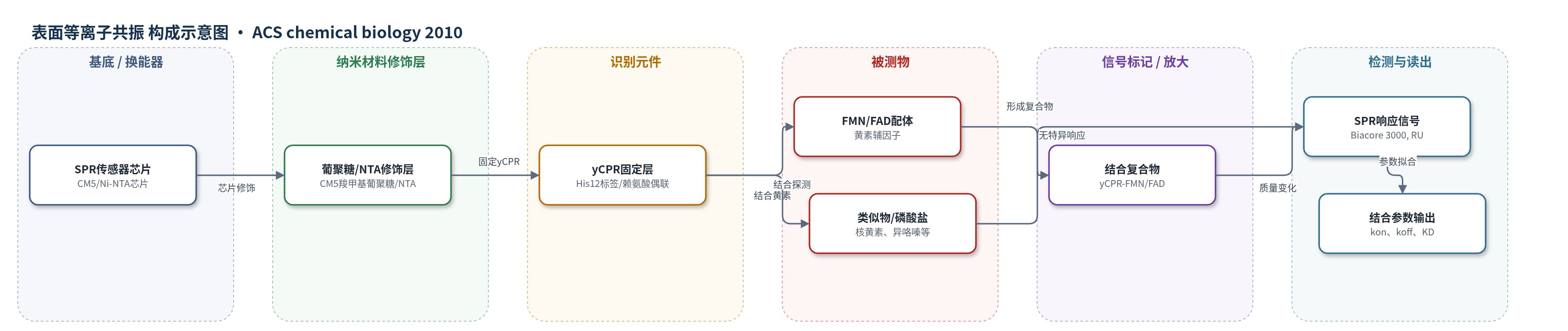
点击图片查看大图 · 依据论文自动绘制
传感器类型
表面等离子共振(SPR)生物传感器
检测对象
FMN(黄素单核苷酸,flavin mononucleotide)、FAD(黄素腺嘌呤二核苷酸,flavin adenine dinucleotide)、黄素类似物(核黄素 riboflavin、二甲基异咯嗪 dimethylalloxazine、异咯嗪 alloxazine、吖啶衍生物等);样品基质为SPR流动池缓冲液(HBS/HBS-EP,含150 mM NaCl和0.005% P20,化合物以DMSO溶解且最终DMSO≤3%)
检测原理
yCPR通过His12标签或赖氨酸共价固定于SPR芯片表面,其表面暴露的FMN2位点作为识别元件。当流动池注入FMN或FAD时,黄素分子与yCPR结合,使芯片表面结合质量增加,局部折射率改变,SPR角/响应单位(RU)随之变化。平衡响应随配体浓度增加并符合单位点结合模型,可计算KD;动力学响应随时间上升和下降,可拟合kon和koff。FMN的磷酸基团与异咯嗪环共同决定特异性,缺少磷酸基团的核黄素、异咯嗪等不产生特异结合;无机磷酸盐和甘油磷酸盐不竞争结合。该方法无酶或标记放大,信号直接来自结合质量变化。
检测灵敏度
原文未报告 LOD、线性范围、灵敏度斜率或相关系数。
效应效果
该SPR系统对黄素辅因子高度选择性:仅FMN和FAD产生化学计量或亚化学计量结合响应,核黄素、二甲基异咯嗪、异咯嗪、氯丙嗪、三氟哌嗪、NADPH/NADP+等无特异正响应;1 mM无机磷酸盐和甘油磷酸盐不干扰结合。CM5表面FMN的KD为(3.0±0.8)×10^-7 M,FAD为(10±3)×10^-6 M,FMN亲和力约高30倍,kon高40–60倍。每组实验至少重复5次,以5次独立测定平均值报告。CM5共价固定可避免NTA表面基线漂移,作者认为结果证明yCPR表面FMN2位点并非结晶假象,支持FMN翻转电子转移机制。
传感器的构成
- 基底/换能器:Biacore SPR传感器芯片(CM5或Ni-NTA),提供表面等离子共振换能界面
- 修饰层:CM5芯片的羧甲基葡聚糖(carboxymethylated dextran)亲水基质,提供羧基用于EDC/NHS偶联;Ni-NTA芯片预固定NTA螯合剂,用于His12标签配位固定
- 识别元件:截短酵母NADPH-细胞色素P450还原酶(yCPR,His12标签),固定于芯片表面,其表面暴露FMN2位点识别FMN/FAD
- 封闭/失活层:乙醇胺(ethanolamine)或HBS-EP缓冲液连续洗涤,失活CM5残余活化羧基,降低非特异结合
- 读出系统:Biacore 3000 SPR仪器,双流通池,以响应单位(RU)记录结合质量变化
中文摘要
NADPH-细胞色素P450还原酶(CPR)通过FAD和FMN将NADPH提供的两个还原当量以单电子转移方式传递给微粒体P450单加氧酶。酵母CPR(yCPR)晶体结构在FMN结合域与连接域界面处发现一个表面暴露的FMN结合位点(FMN2),此外还有大鼠CPR中观察到的埋藏位点。该发现为CPR分子内(FAD与FMN之间)和分子间(FMN与P450之间)电子转移提供了可检验假说。为验证FMN2位点占据并非结晶假象,作者采用表面等离子共振(SPR)生物传感器技术,在不同传感器芯片表面固定yCPR,并用FMN、FAD、核黄素、二甲基异咯嗪、异咯嗪及类似异咯嗪环的小分子进行动力学和平衡结合实验。结果显示只有FMN和FAD产生化学计量结合响应;FMN结合亲和力为亚微米摩尔级,比FAD高约30倍;yCPR/FMN复合物的结合速率常数比yCPR/FAD高至多60倍。结果表明该表面暴露位点对黄素具有高度选择性,FMN结合更受青睐,且FMN的磷酸基团和异咯嗪环均为结合所必需。
英文摘要
NADPH-cytochrome P450 reductase (CPR) transfers two reducing equivalents derived from NADPH via FAD and FMN to microsomal P450 monooxygenases in one-electron transfer steps. The crystal structure of yeast CPR (yCPR) contains a surface-exposed FMN binding site (FMN2 site) at the interface of the FMN binding and connecting domains, in addition to the single buried site that has been observed in rat CPR. This finding provides a testable hypothesis of how intramolecular (between FAD and FMN) and intermolecular (between FMN and P450) electron transfer may occur in CPR. To verify that occupancy of the FMN2 site is not an artifact of crystallization, a surface plasmon resonance (SPR) biosensor technique has been applied to probe the selectivity of this site under functional conditions. A series of kinetic and equilibrium binding experiments involving yCPR immobilized on different sensor chip surfaces was performed using FMN and FAD, as well as FMN-derived compounds, including riboflavin, dimethylalloxazine, and alloxazine, and other molecules that resemble the planar isoalloxazine ring structure. Only FMN and FAD showed stoichiometric binding responses. Binding affinity for FMN was in the submicromolar range, 30 times higher than that for FAD. Association kinetic rates for the yCPR/FMN complex were up to 60-fold higher than for the yCPR/FAD complex. Taken together, these data indicate that (i) the surface-exposed site in yCPR is highly selective toward binding flavins, (ii) binding of FMN in this site is notably favored, and finally, (iii) both the phosphate group and the isoalloxazine ring of FMN are essential for binding.