组成图示
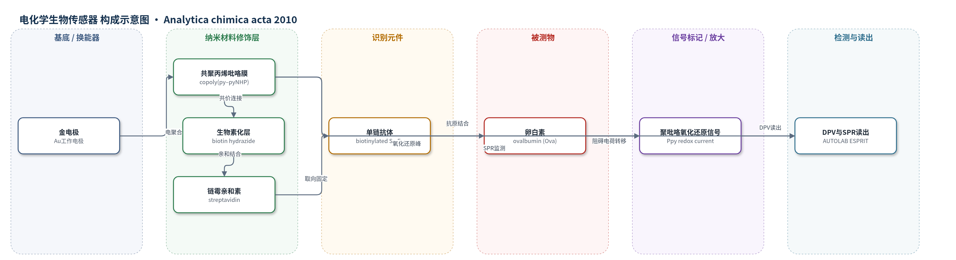
点击图片查看大图 · 依据论文自动绘制
传感器类型
电化学生物传感器
检测对象
卵白素(ovalbumin, Ova)、特异性抗原(specific antigen);样品基质:PBS缓冲液(10 mM, pH 7.4)
检测原理
传感器以金电极为基底,在其上电聚合copoly(py–pyNHP)导电聚合物膜。pyNHP上的N-羟基邻苯二甲酰亚胺酯与生物素酰肼氨基共价连接,再经链霉亲和素-生物素体系定向固定生物素化Sc-Fv抗体。抗原(Ova)与抗体结合后形成免疫复合物,使聚吡咯膜表面电荷传输和掺杂离子渗透受阻,导致0.16 V/SCE处聚吡咯氧化峰电流随抗原浓度增加而下降。SPR则通过界面质量/折射率变化引起共振角增加来同步监测结合事件。该体系为无标记检测,信号放大依赖DPV对聚吡咯法拉第氧化还原电流的高灵敏读取,而非酶催化或核酸扩增。
检测灵敏度
LOD: 1 pg mL−1(原文:a low concentration of 1 pg mL−1 was detected);线性范围: 1 pg mL−1–100 ng mL−1;灵敏度: 17.6 nA (ng mL)−1
效应效果
该传感器在PBS中检测Ova,校准曲线在1 pg mL−1至100 ng mL−1范围内呈线性,灵敏度为17.6 nA (ng mL)−1,3次测量相对标准偏差为5%。SPR显示再生后信号可回到基线,表明稳定性良好;不同分析间响应可重复。以人IgG作为非特异性蛋白测试时,仅出现溶液折射率变化引起的短暂角度上升,孵育后角度下降,说明无明显非特异性固定,选择性较好。作者认为该无标记聚吡咯免疫传感器结合DPV放大和生物素/链霉亲和素定向固定,可推广至多种抗体-抗原体系,具有微型化和临床/药物发现应用潜力。
传感器的构成
- 基底/换能器电极:金电极(Au)作为工作电极和SPR传感芯片金属层,配合Ag/AgCl参比电极与Pt对电极构成三电极体系
- 导电聚合物修饰层:共聚丙烯吡咯(copoly(py–pyNHP)),由吡咯(py)和3-N-羟基邻苯二甲酰亚胺酯吡咯(pyNHP)电聚合形成,py作间隔单元,pyNHP提供活化酯连接位点
- 生物素连接层:生物素酰肼(biotin hydrazide)与pyNHP活化酯共价连接,形成copoly(py–pyBiotin),提供生物素结合位点
- 亲和固定层:链霉亲和素(streptavidin)通过生物素-链霉亲和素高亲和作用固定抗体并控制抗体取向
- 识别元件:生物素化单链抗体片段(biotinylated Sc-Fv Ab)识别特异性抗原
- 封闭剂:酪蛋白(casein)封闭自由结合位点,减少非特异性结合
- 信号标记物:无标记(label-free),聚吡咯自身氧化还原峰作为电化学信号来源
中文摘要
本文报道了一种基于生物素化单链可变片段(Sc-Fv)抗体固定在共聚丙烯吡咯膜上的无标记电化学生物传感器。该免疫传感器通过生物素/链霉亲和素体系,将生物素化单链抗体固定在电聚合聚吡咯共聚物膜上,首次实现了高效抗原检测。传感器响应通过表面等离子共振(SPR)和差分脉冲伏安法(DPV)对聚吡咯氧化还原信号的电化学分析进行监测。共聚物由吡咯(py)作为间隔单元和3位带N-羟基邻苯二甲酰亚胺酯基团的吡咯(pyNHP)作为生物分子固定连接剂组成,其py:pyNHP比例针对抗体固定和抗原检测进行了优化。SPR实时监测电聚合过程及传感器逐步构建;FT-IR证实共聚物组成和生物分子共价连接效率;SEM分析膜形貌。结果表明,优化共聚物组成可获得有序抗体层,并产生强而高的聚吡咯氧化还原信号。SPR和DPV均证实特异性抗原检测,通过测量聚吡咯氧化还原信号变化可检测低至1 pg mL−1的抗原。
英文摘要
An electrochemical label-free immunosensor based on a biotinylated single-chain variable fragment (Sc-Fv) antibody immobilized on copolypyrrole film is described. An efficient immunosensor device formed by immobilization of a biotinylated single-chain antibody on an electropolymerized copolymer film of polypyrrole using biotin/streptavidin system has been demonstrated for the first time. The response of the biosensor toward antigen detection was monitored by surface plasmon resonance (SPR) and electrochemical analysis of the polypyrrole response by differential pulse voltammetry (DPV). The composition of the copolymer formed from a mixture of pyrrole (py) as spacer and a pyrrole bearing a N-hydroxyphthalimidyl ester group on its 3-position (pyNHP), acting as agent linker for biomolecule immobilization, was optimized for an efficient immunosensor device. The ratio of py:pyNHP for copolymer formation was studied with respect to the antibody immobilization and antigen detection. SPR was employed to monitor in real time the electropolymerization process as well as the step-by-step construction of the biosensor. FT-IR demonstrates the chemical copolymer composition and the efficiency of the covalent attachment of biomolecules. The film morphology was analyzed by electron scanning microscopy (SEM). Results show that a well organized layer is obtained after Sc-Fv antibody immobilization thanks to the copolymer composition defined with optimized pyrrole and functionalized pyrrole leading to high and intense redox signal of the polypyrrole layer obtained by the DPV method. Detection of specific antigen was demonstrated by both SPR and DPV, and a low concentration of 1 pg mL(-1) was detected by measuring the variation of the redox signal of polypyrrole.