组成图示
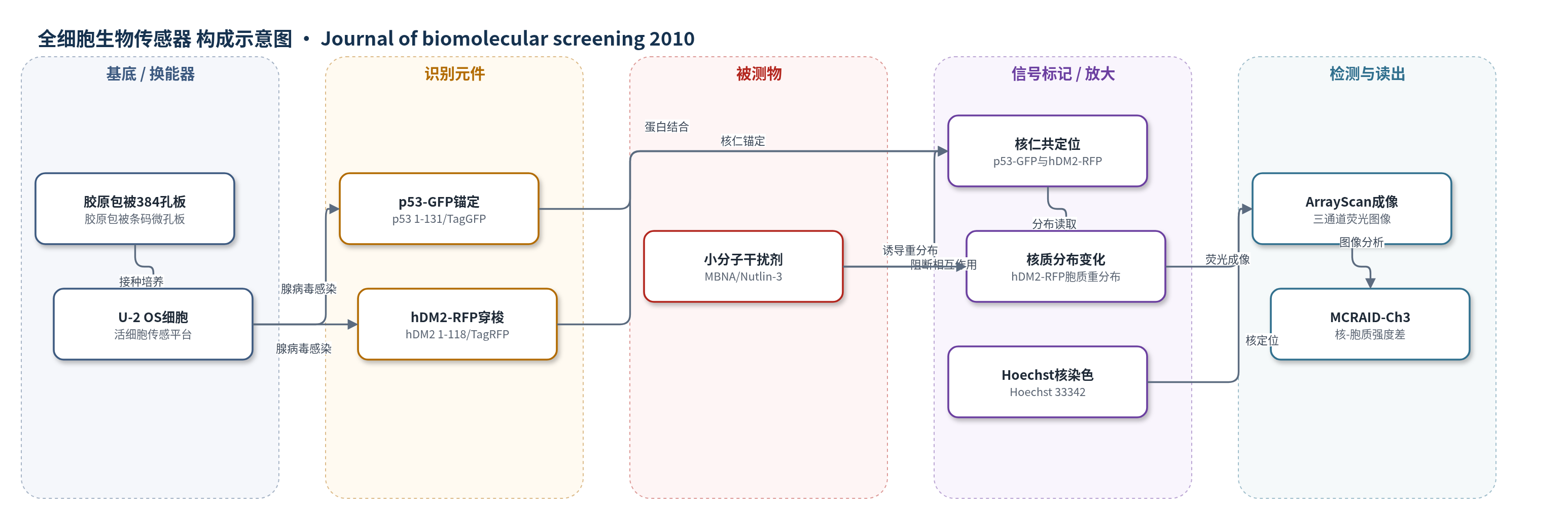
点击图片查看大图 · 依据论文自动绘制
传感器类型
全细胞生物传感器
检测对象
p53-hDM2蛋白-蛋白相互作用(p53-hDM2 PPI)及其小分子干扰剂(protein-protein interaction disruptors, PPIDs;MBNA、Nutlin-3);样品基质:NIH小分子化合物库(DMSO/McCoy's 5A培养基,384孔板)
检测原理
该传感器以活细胞为换能平台。U-2 OS细胞共感染两种腺病毒构建体:p53 1-131与monomeric TagGFP融合并含NLS,被锚定在核仁;hDM2 1-118与monomeric TagRFP融合并含NLS/NES,可在核质间穿梭。无干扰剂时,hDM2-RFP与p53-GFP结合后被锚定在核仁,核内RFP平均荧光强度高于胞质。加入p53-hDM2相互作用干扰剂后,hDM2-RFP与p53-GFP解离,经NES输出至胞质,导致核-胞质RFP平均强度差MCRAID-Ch3下降。Hoechst 33342染色核酸以生成核掩膜,ArrayScan VTI采集Hoechst、GFP、RFP三通道图像,图像分析计算MCRAID-Ch3并以DMSO和Nutlin-3对照归一化,得到相互作用抑制率。信号随干扰剂浓度升高而降低,无额外化学放大,依赖荧光蛋白定位与多参数图像分析。
检测灵敏度
未报告LOD、线性范围、灵敏度斜率或相关系数;IC50: 30–50 µM(原文:IC50s between 30 and 50 µM);Nutlin-3 ~30-fold more potent
效应效果
该PPIB在697块384孔板中平均Z′因子0.64±0.13,S/B 31.0±9.8倍,稳健可重复。初筛220,017个化合物,312个(0.14%)≥50%抑制;确认后3个MBNA可重复干扰p53-hDM2,IC50 30–50 µM。多参数图像分析排除133个细胞毒性(0.06%)和3037个荧光伪影(1.38%)。HCT116-WT中,MBNAs升高p53、hDM2、p21,诱导G1阻滞,凋亡率由6%升至37%(SID 17433115),低于Nutlin-3的58%;生长抑制IC50约3.9 µM,与Nutlin-3的3.7 µM相当。MBNAs在p53缺失细胞中凋亡升至22.9%,并在p53/p21缺失细胞系保持约4 µM生长抑制IC50,显示部分p53非依赖性。
传感器的构成
- 培养基底:胶原包被384孔条码微孔板(collagen-coated barcoded microplate, Greiner Bio-One #781986),承载U-2 OS细胞并适配384孔HCS。
- 传感细胞:U-2 OS细胞,经两种重组腺病毒共感染,作为活细胞蛋白-蛋白相互作用传感平台。
- 锚定识别元件:p53 1-131/monomeric TagGFP融合蛋白(p53-GFP),含核定位序列(NLS),定位于核仁并锚定hDM2-RFP。
- 穿梭识别/报告元件:hDM2 1-118/monomeric TagRFP融合蛋白(hDM2-RFP),含NLS和核输出序列(NES),作为可核质穿梭的相互作用伙伴。
- 荧光标记物:monomeric TagGFP和monomeric TagRFP(Evrogen),分别标记p53和hDM2,提供GFP/RFP荧光信号。
- 核定位标记:Hoechst 33342,染色核酸,用于聚焦、定义核掩膜并区分核/胞质区域。
- 固定与成像介质:7.4%甲醛(formaldehyde)和PBS,固定细胞并保留荧光蛋白亚细胞分布,供ArrayScan VTI成像。
- 读出平台:ArrayScan VTI自动成像平台,10× 0.3 NA物镜与XF93滤片组,采集Hoechst/GFP/RFP三通道图像。
中文摘要
本文报道了一种新型p53-hDM2蛋白-蛋白相互作用生物传感器(PPIB)在高内涵筛选(HCS)中的应用。作者利用U-2 OS细胞共感染表达p53-GFP和hDM2-RFP的重组腺病毒构建体,使p53-GFP锚定于核仁,hDM2-RFP作为可核质穿梭的相互作用伙伴;当小分子干扰p53-hDM2结合时,hDM2-RFP重分布至胞质,通过三通道荧光成像和图像分析定量。该PPIB在筛选美国国立卫生研究院(NIH)220,017个化合物时表现出稳健性和可重复性,多参数图像分析可排除细胞毒性和荧光伪影。筛选获得3个结构相关的甲基苯并萘啶-5-胺(MBNA)化合物,在PPIB中IC50为30–50 µM。在野生型p53的HCT116细胞中,MBNAs升高p53蛋白水平,上调p53靶基因,诱导G1期阻滞、凋亡并抑制增殖,IC50约4 µM。Nutlin-3在PPIB中效力更强,但细胞生物学效应相当;MBNAs还能在p53缺失细胞中增加凋亡,并在p53或p21缺失同基因细胞系中保持相似生长抑制效力。
英文摘要
In recent years, advances in structure-based drug design and the development of an impressive variety of high-throughput screening (HTS) assay formats have yielded an expanding list of protein-protein interaction inhibitors. Despite these advances, protein-protein interaction targets are still widely considered difficult to disrupt with small molecules. The authors present here the results from screening 220,017 compounds from the National Institute of Health's small-molecule library in a novel p53-hDM2 protein-protein interaction biosensor (PPIB) assay. The p53-hDM2 positional biosensor performed robustly and reproducibly throughout the high-content screening (HCS) campaign, and analysis of the multiparameter data from images of the 3 fluorescent channels enabled the authors to identify and eliminate compounds that were cytotoxic or fluorescent artifacts. The HCS campaign yielded 3 structurally related methylbenzo-naphthyridin-5-amine (MBNA) hits with IC(50)s between 30 and 50 microM in the p53-hDM2 PPIB. In HCT116 cells with wild-type (WT) p53, the MBNAs enhanced p53 protein levels, increased the expression of p53 target genes, caused a cell cycle arrest in G1, induced apoptosis, and inhibited cell proliferation with an IC(50) ~4 microM. The prototype disruptor of p53-hDM2 interactions Nutlin-3 was more potent than the MBNAs in the p53-hDM2 PPIB assay but produced equivalent biological results in HCT116 cells WT for p53. Unlike Nutlin-3, however, MBNAs also increased the percentage of apoptosis in p53 null cells and exhibited similar potencies for growth inhibition in isogenic cell lines null for p53 or p21. Neither the MBNAs nor Nutin-3 caused cell cycle arrest in p53 null HCT116 cells. Despite the relatively modest size of the screening library, the combination of a novel p53-hDM2 PPIB assay together with an automated imaging HCS platform and image analysis methods enabled the discovery of a novel chemotype series that disrupts p53-hDM2 interactions in cells.