组成图示
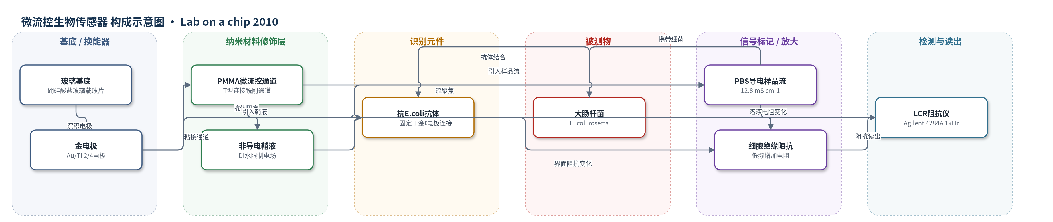
点击图片查看大图 · 依据论文自动绘制
传感器类型
微流控生物传感器
检测对象
大肠杆菌(Escherichia coli, E. coli);样品基质为 PBS 磷酸盐缓冲液(导电样品流),鞘液为去离子水
检测原理
该传感器基于微流控流聚焦与电化学阻抗测量。改进T型通道中,非导电去离子水鞘液从顶部和侧面包裹导电PBS样品流,形成二维聚焦层,使交流电场主要限制在样品流内。金电极施加10 mV、1 kHz小信号,4电极配置分离电流与传感电极,降低双电层阻抗,使低频溶液电阻可测。金电极表面固定抗E. coli抗体,大肠杆菌结合后,由于细胞膜在低频呈绝缘性,会阻挡电流路径,使有效溶液电阻增加、有效电极面积和电容下降。提高鞘液-样品流量比可减小聚焦层截面积并增强电场限制,但鞘液与样品界面扩散使导电离子进入鞘液,抬高基线电阻并抵消细胞绝缘信号;提高流速可缩短停留时间、减轻扩散。因此信号随结合细胞量增加表现为低频阻抗/电阻上升,但需同时优化流量比与流速。
检测灵敏度
原文未报告 LOD、线性范围、灵敏度斜率或 R^2。
效应效果
实验重复3次并给出标准差。4电极配置在FRR 50:2时电阻由31.5±0.7 kΩ升至38.1±1.0 kΩ,ΔR=20.8%;电容由1.36±0.02 nF降至1.31±0.02 nF,ΔC=-3.7%。2电极配置在FRR 50:2时电阻ΔR=13.5%,电容ΔC=-13.9%,对电极界面变化更敏感。随着FRR增至100:2和200:2,4电极ΔR为15.6%和14.8%,2电极ΔR降至11.2%和8.0%,说明高流量比下鞘液-样品扩散削弱信号。BSA封闭降低非特异结合;作者认为100:2 mL/min(FRR=50,Re=2.7)是该设计的较优条件。
传感器的构成
- 基底/换能器电极:硼硅酸盐玻璃载玻片(borosilicate glass microscope slides)作为基底,表面光刻制备金电极(Au 300 nm)和钛粘附层(Ti 30 nm),构成2/4电极阻抗换能器。
- 识别元件:山羊抗大肠杆菌抗体(goat anti-E. coli antibody),经sulfo-LC-SPDP和DTT处理后直接固定于金电极表面,用于特异性捕获E. coli。
- 微流控通道:PMMA(聚甲基丙烯酸甲酯)铣削通道,含鞘液入口、样品入口和聚焦通道,改进T型连接,用UV固化胶粘接到玻璃电极片,形成二维流聚焦虚拟通道。
- 鞘液层:去离子水(DI water,非导电,电导率0.08 mS cm-1),从顶部和侧面包裹样品流,将电场限制在导电聚焦层内。
- 样品导电层:PBS(磷酸盐缓冲液,电导率12.8 mS cm-1),作为导电样品流携带大肠杆菌,形成被聚焦的阻抗测量介质。
- 封闭剂:BSA(牛血清白蛋白)流经通道,用于减少非特异性结合。
- 信号标记物:无外加电化学标记物;E. coli rosetta细胞本身在低频下因细胞膜绝缘而改变电极界面与溶液阻抗。
- 读出系统:Agilent 4284A LCR meter,施加10 mV、1 kHz交流信号,进行2/4电极阻抗测量,并用LabVIEW控制注射泵和采集数据。
中文摘要
本文研究了阻抗型生物传感器中非导电鞘液与导电样品流之间扩散对测量的影响。采用2电极和4电极配置进行阻抗测量;4电极设计将电流电极与传感电极分离,可避免双电层阻抗的有害影响,使低频(<1 kHz)阻抗测量成为可能。器件采用改进T型连接微流控通道,样品通道截面小于聚焦通道,仅用一路鞘液即可实现样品流的二维流聚焦。通过选择去离子水作为非导电鞘液、磷酸盐缓冲液作为导电样品流,将电场限制在聚焦样品流内。作者对系统的电学参数和流动参数进行了表征,考察流速和鞘液-样品流量比对聚焦流的影响。有限元模拟和共聚焦显微镜证实,提高流量比可减小聚焦流截面积。抗体介导的大肠杆菌与电极表面结合后,低频溶液电阻增加。结果表明,鞘液与样品界面处的扩散传质限制了流聚焦带来的灵敏度提升;提高流速可部分抵消扩散效应。为优化检测灵敏度,需同时考虑流动参数与传质,在减小聚焦流截面积的同时抑制导电离子向鞘液扩散。
英文摘要
This paper investigated the effects of diffusion between non-conductive sheath and conductive sample fluids in an impedance-based biosensor. Impedance measurements were made with 2- and 4-electrode configurations. The 4-electrode design offers the advantage of impedance measurements at low frequencies (<1 kHz) without the deleterious effects of double layer impedance which are present in the 2-electrode design. Hydrodynamic flow focusing was achieved with a modified T-junction design with a smaller cross-section for the sample channel than for the focusing channel, which resulted in 2D focusing of the sample stream with just one sheath stream. By choosing a non-conductive sheath fluid and a conductive sample fluid, the electric field was confined to the focused stream. In order to utilize this system for biosensing applications, we characterized it for electrical and flow parameters. In particular, we investigated the effects of varying flow velocities and flow-rate ratios on the focused stream. Increasing flow-rate ratios reduced the cross-sectional area of the focused streams as was verified by finite element modeling and confocal microscopy. Antibody mediated binding of Escherichia coli to the electrode surface caused an increase in solution resistance at low frequencies. The results also showed that the diffusion mass transport at the interface of the two streams limited the benefits of increased flow focusing. Increasing flow velocities could be used to offset the diffusion effect. To optimize detection sensitivity, flow parameters and mass transport must be considered in conjunction, with the goal of reducing diffusion of conducting species out of the focused stream while simultaneously minimizing its cross-sectional area.