组成图示
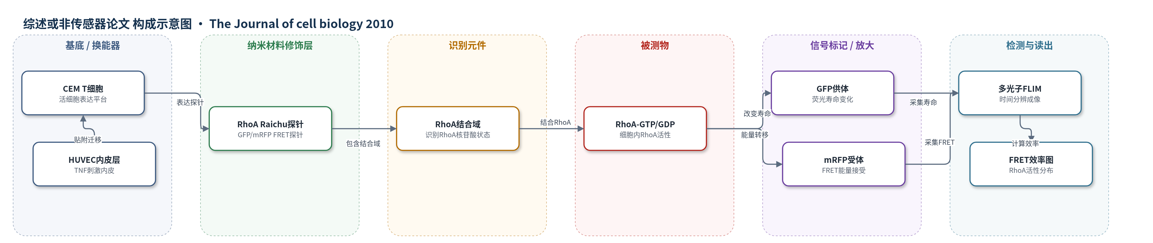
点击图片查看大图 · 依据论文自动绘制
传感器类型
综述或非传感器论文
检测对象
RhoA 活性(RhoA activity,GTP/GDP 负载状态);样品基质:活 CEM T 细胞或 T 淋巴细胞在 TNF 刺激的人脐静脉内皮细胞(HUVEC)上爬行/跨内皮迁移
检测原理
RhoA Raichu 探针由 RhoA 结合域、GFP 供体和 mRFP 受体组成,在 CEM T 细胞内表达。当内源 RhoA 结合探针并结合 GTP 或 GDP 时,探针构象改变,使 GFP 与 mRFP 的距离或取向发生变化,从而改变 FRET 效率。多光子 FLIM 测量 GFP 荧光寿命,FRET 效率定义为 1 减去供体加受体寿命与单独供体寿命之比;RhoA 活性升高时 FRET 效率/寿命信号改变。通过逐帧成像,可定位前缘、尾足和板状区中 RhoA 活性的时空变化,并关联膜伸出、回缩与尾足收缩。
检测灵敏度
—
效应效果
筛选显示 RhoA 缺失对 TEM 抑制最强,且抑制程度与敲低效率相关;RhoA 缺失细胞完成 TEM 时间延迟。约 75±4% 的迁移细胞采用细胞间途径,18±3% 的贴附细胞发生 TEM。FLIM 定量显示运动细胞前缘 RhoA 活性峰值高于侧缘(P=0.04),尾足高于侧缘(P=0.01)。前缘离散 RhoA 激活事件中,52±7% 随后发生膜伸出,36±4% 发生回缩,12±3% 未立即关联。探针表达未明显影响膜动力学、极性或 TEM,FRET 效率不依赖探针浓度,显性负突变探针 FRET 效率更低,支持信号特异性。
传感器的构成
- 探针识别层:RhoA Raichu 探针(含 RhoA 结合域)结合内源 RhoA,报告其 GTP/GDP 负载状态
- 荧光供体层:GFP 作为 FRET 供体,其荧光寿命随 RhoA 活性变化
- 荧光受体层:mRFP 作为 FRET 受体,与 GFP 组成 FRET 对
- 表达基质:CEM T 细胞或 T 淋巴细胞,作为活细胞表达与成像平台
- 内皮微环境:TNF 刺激的 HUVEC,提供 T 细胞贴附、爬行与 TEM 基质
- 读出系统:多光子 FLIM/FRET 显微镜,采集 GFP 寿命并计算 FRET 效率
中文摘要
跨内皮迁移(TEM)是白细胞从血管进入组织的受调控过程,Rho GTP 酶参与该过程,但各成员贡献不明。本研究用 RNA 干扰筛选鉴定影响 T 细胞 TEM 的 Rho GTP 酶,证明 RhoA 对该过程至关重要。RhoA 缺失导致迁移极性丧失:细胞缺乏前缘和尾足结构,转而形成稳定的窄突起,且突起与收缩事件去极化分布。通过成像 RhoA 活性生物传感器,发现 RhoA 在迁移和跨内皮迁移 T 细胞的前缘局部、动态激活,其激活先于膜伸出和回缩事件;在尾足中,RhoA 激活与 ROCK 介导的收缩相关。Rho 鸟苷酸交换因子 GEF-H1 促进尾足收缩,但不影响前缘。结果表明,RhoA 活性在 T 细胞前后端受到动态调控,以协调 TEM。
英文摘要
Transendothelial migration (TEM) is a tightly regulated process whereby leukocytes migrate from the vasculature into tissues. Rho guanosine triphosphatases (GTPases) are implicated in TEM, but the contributions of individual Rho family members are not known. In this study, we use an RNA interference screen to identify which Rho GTPases affect T cell TEM and demonstrate that RhoA is critical for this process. RhoA depletion leads to loss of migratory polarity; cells lack both leading edge and uropod structures and, instead, have stable narrow protrusions with delocalized protrusions and contractions. By imaging a RhoA activity biosensor in transmigrating T cells, we find that RhoA is locally and dynamically activated at the leading edge, where its activation precedes both extension and retraction events, and in the uropod, where it is associated with ROCK-mediated contraction. The Rho guanine nucleotide exchange factor (GEF) GEF-H1 contributes to uropod contraction but does not affect the leading edge. Our data indicate that RhoA activity is dynamically regulated at the front and back of T cells to coordinate TEM.