组成图示
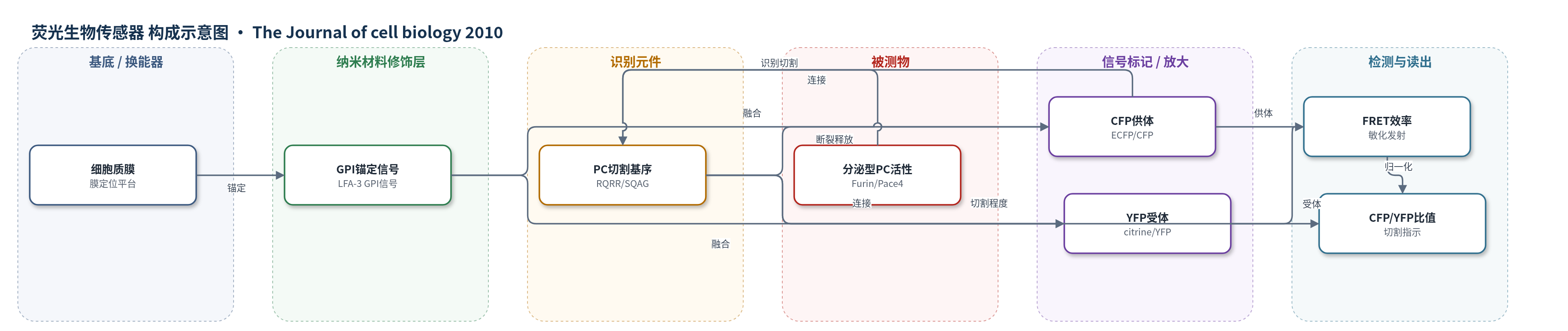
点击图片查看大图 · 依据论文自动绘制
传感器类型
荧光生物传感器
检测对象
前蛋白转化酶(proprotein convertases, PCs)活性,包括Furin、Pace4、Pcsk5、PC7;样品基质为活细胞(HEK293T、LoVo、小鼠胚胎干细胞ES)、小鼠囊胚/植入胚胎组织及成体组织切片。
检测原理
CLIP由CFP、PC识别基序RQRR和YFP串联组成,并通过GPI锚定在质膜。分泌型PC(如Furin、Pace4)识别RQRR并切割连接肽,使CFP片段从膜上释放,而YFP-GPI仍保留在膜上。切割前后CFP与YFP距离改变,导致FRET效率下降;未切割时CFP激发能量转移至YFP,产生敏化发射。检测时用校正FRET信号除以YFP信号得到NFRET,PC活性越高,切割越多,NFRET越低,CFP/YFP比值越高。加入α1-PDX或CMK可抑制切割,使NFRET升高。该传感器为比率型荧光读出,无需外源标记或酶放大。
检测灵敏度
原文未报告LOD、线性范围、灵敏度斜率或相关系数。
效应效果
CLIP特异性较高:生理浓度凝血酶(<2 U/ml)无影响,α1-PDX或CMK可阻断切割;Furin/Pace4双敲除ES细胞中NFRET接近CLIPm,外源Furin、Pcsk5B或PC7可恢复,PC1/3、PC2、PC4不能。0.4 µm滤膜共培养证明可检测非接触旁分泌活性。定量FRET显示CLIPm NFRET为30±4%,CMK后41±4%或37±6%,光漂白YFP后CFP增加34±4%,CMK后NFRET约10%/h升高。囊胚CFP/YFP在DKO为0.84±0.13,对照0.21±0.04;E6.5 DKO约2倍、CLIPm约5倍;离体DKO 5–7 h内CFP/YFP升50%。作者称其为体内检测PC活性的新工具。
传感器的构成
- 基底/定位平台:细胞质膜(plasma membrane, PM),作为GPI锚定CLIP/CLIPm的界面。
- 表达定位层:乳酸酶-氟苷水解酶信号序列(ss)与GPI锚定信号(LFA-3 GPI),将CLIP/CLIPm靶向质膜。
- 供体荧光蛋白层:ECFP(CFP),作为FRET供体,被PC切割后从膜上释放。
- 识别切割层:PC共识切割基序RQRR,连接CFP与YFP,被Furin/Pace4等PC识别并切割;阴性对照CLIPm使用抗切割序列SQAG。
- 受体荧光蛋白层:citrine(YFP),作为FRET受体并保留在质膜,用于归一化膜上生物传感器总量。
- 换能读出层:FRET荧光共振能量转移,CFP激发下YFP敏化发射,切割导致FRET降低。
- 转基因表达元件:鸡β-actin/CMV杂交启动子、β-actin第一内含子和兔β-globin 3' UTR,用于小鼠体内表达。
中文摘要
哺乳动物中,前蛋白转化酶(proprotein convertases, PCs)家族由9种丝氨酸蛋白酶组成,其中Furin、Pace4、Pcsk5和PC7广泛表达,可切割多种生长因子、受体、黏附分子、神经肽、金属蛋白酶、病毒包膜糖蛋白及细菌内毒素的前体。然而,经典遗传学方法难以阐明各PC在正常组织和疾病中的具体作用。作者此前发现Furin和Pace4在早期胚胎发生中共同激活TGF-β相关Nodal前体,但二者何时何地开始活性尚不清楚。为此,作者开发了一种新型细胞膜靶向荧光生物传感器——细胞表面连接蛋白水解指示剂(CLIP)。在野生型及Furin/Pace4缺陷胚胎干细胞和胚胎中活体成像显示,Furin和Pace4在囊胚阶段的内细胞团中已具有活性,并能以细胞自主和非自主方式切割膜结合底物。CLIP还在植入胚胎的上胚层中被切割,部分依赖于子宫中一种独立于合子Furin和Pace4的新型活性,提示母源PC参与胚胎发育。CLIP具有前所未有的灵敏度和空间分辨率,为阐明PC体内功能提供新工具。
英文摘要
Axis formation and allocation of pluripotent progenitor cells to the germ layers are governed by the TGF-β-related Nodal precursor and its secreted proprotein convertases (PCs) Furin and Pace4. However, when and where Furin and Pace4 first become active have not been determined. To study the distribution of PCs, we developed a novel cell surface-targeted fluorescent biosensor (cell surface-linked indicator of proteolysis [CLIP]). Live imaging of CLIP in wild-type and Furin- and Pace4-deficient embryonic stem cells and embryos revealed that Furin and Pace4 are already active at the blastocyst stage in the inner cell mass and can cleave membrane-bound substrate both cell autonomously and nonautonomously. CLIP was also cleaved in the epiblast of implanted embryos, in part by a novel activity in the uterus that is independent of zygotic Furin and Pace4, suggesting a role for maternal PCs during embryonic development. The unprecedented sensitivity and spatial resolution of CLIP opens exciting new possibilities to elucidate PC functions in vivo.