组成图示
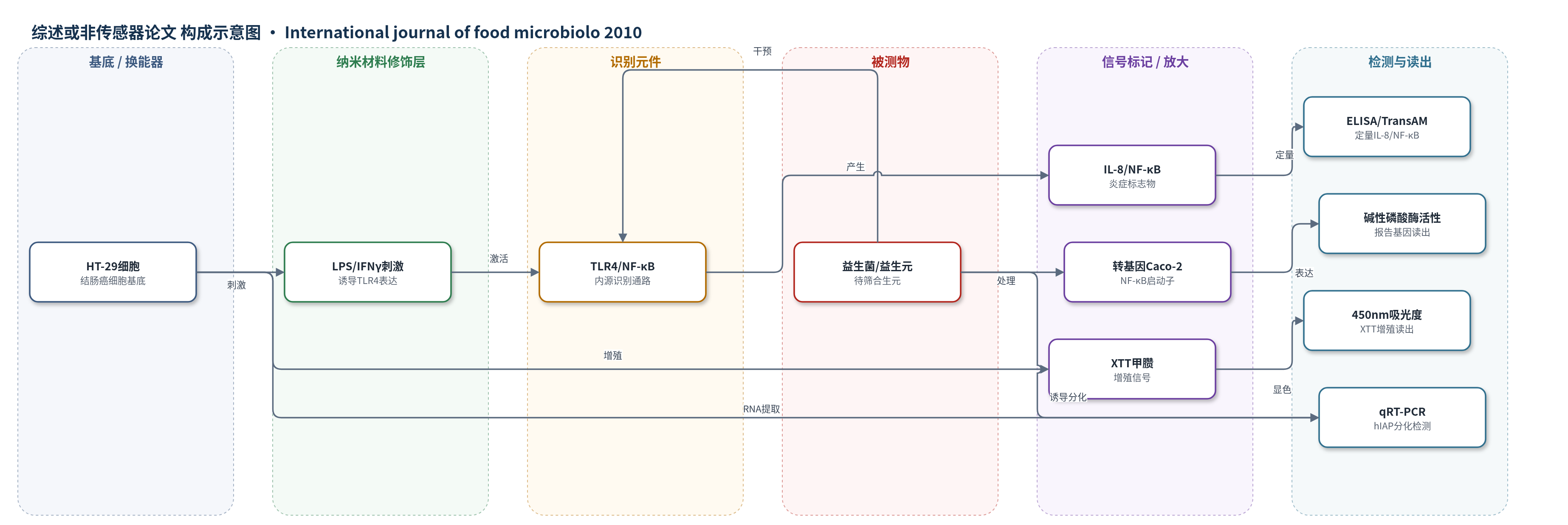
点击图片查看大图 · 依据论文自动绘制
传感器类型
综述或非传感器论文
检测对象
益生菌乳酸菌(probiotic lactic acid bacteria)、低聚葡萄糖(glucooligosaccharides, GOS)、合生元(synbiotics);样品基质:体外细胞培养体系(HT-29/Caco-2细胞培养上清、核提取物)
检测原理
本文并非传统传感器,而是基于细胞模型的体外检测。IFNγ先上调HT-29细胞表面TLR4表达,LPS结合TLR4后激活NF-κB信号通路,诱导IL-8分泌;加入益生菌、益生元或合生元后,若其具有抗炎作用,则IL-8和核内NF-κB水平下降,经ELISA/TransAM试剂盒定量。转基因Caco-2细胞中,NF-κB诱导型启动子驱动碱性磷酸酶表达,酶活性与NF-κB激活程度相关,用于二次验证。增殖检测中,活细胞线粒体脱氢酶将XTT还原为有色甲臜,450 nm吸光度与细胞数量/增殖程度成正比;合生元抑制增殖并上调hIAP,qRT-PCR以β-actin内参定量,提示分化机制。
检测灵敏度
原文未报告 LOD、线性范围、灵敏度斜率或相关系数。
效应效果
筛选显示11株乳酸菌在HT-29模型中具抗炎活性,其中5株同时显著抑制IL-8分泌和NF-κB激活;转基因Caco-2报告基因模型进一步确认多数有效菌株。益生元GOS单独无抗炎作用,与菌联合未增强抗炎效果。抗增殖方面,单菌抑制有限且重现性差,B. breve R0070/L. lactis R1058/OA合生元抑制率最高,为66.31%±2.05%,讨论中称可达约70%。剂量实验表明MOI 100和OA 10 g/L有效,MOI低于10或OA降至0.1 g/L时作用减弱。hIAP表达上调2倍,与14天分化对照的2.5倍无显著差异。作者认为该合生元具抗炎和抗增殖潜力,需动物模型验证。
传感器的构成
- 非传感器体系:本文无传感器结构,采用体外细胞检测体系
- 细胞基底:HT-29人结肠癌细胞,提供炎症与增殖响应平台
- 刺激/识别元件:LPS与IFNγ,诱导TLR4表达并激活NF-κB通路
- 信号标记物:分泌型IL-8与核内激活NF-κB,作为炎症生物标志物
- 读出方法:ELISA与TransAM NF-κB试剂盒,定量IL-8和NF-κB
- 报告基因系统:转基因Caco-2细胞,NF-κB启动子控制碱性磷酸酶
- 增殖检测:XTT被线粒体脱氢酶还原为甲臜,450 nm吸光度
- 分化检测:实时RT-PCR检测hIAP与β-actin mRNA
中文摘要
本文在体外模型中筛选益生菌、益生元和合生元的抗炎与抗增殖作用。为模拟炎症性肠病对革兰阴性菌的反应,先用IFNγ上调HT-29细胞TLR4表达,再以LPS刺激,检测分泌型IL-8和激活型NF-κB。结果显示,11株乳酸菌具有抗炎活性,其中5株显著抑制IL-8分泌和NF-κB激活;益生元低聚葡萄糖单独无抗炎作用。随后用NF-κB启动子控制报告基因的转基因Caco-2细胞验证,确认部分菌株的抑制作用。HT-29癌细胞增殖用XTT法检测,仅3株菌单独抑制增殖且重现性有限;双联或三联益生菌与低聚葡萄糖联合可显著降低增殖,其中短双歧杆菌R0070、乳乳球菌R1058与低聚交替糖OA的合生元抑制率最高,达66.31%±2.05%。剂量实验表明细菌和OA均参与作用。实时RT-PCR显示该合生元上调肠碱性磷酸酶hIAP表达,提示其通过诱导细胞分化抑制增殖。
英文摘要
There is emerging evidence of the efficiency of probiotic, prebiotic and synbiotic treatments in inflammatory bowel diseases (IBDs) and one of their long-term complications, colorectal cancer (CRC). In this study, various strains of probiotic lactic acid bacteria, prebiotic glucooligosaccharides (GOS) or a synbiotic combination of the two were screened for anti-inflammatory and anti-proliferative effects in different in vitro models in the context of such diseases. To mimic IBD response to Gram negative bacteria, HT-29 cells were sensitised to inflammatory response to lipopolysaccharide (LPS) by IFNγ which increased expression of TLR4, the LPS biosensor, and were then treated by probiotics, prebiotics and synbiotics. Secreted IL-8 and activated NF-κB were monitored as inflammation biomarkers. A selection of active strains were then subjected to a second inflammatory cell culture model consisting of inflammatory activated transgenic Caco-2 cells transfected by a reporter gene under the control of NF-κB inducible promoter. Quantification of reporter gene expression allowed us to demonstrate some probiotic inhibitory properties or to confirm such characteristics in two different models. Proliferation of cancerous HT-29 cells was monitored by XTT assay. Only three probiotic strains induced a proliferation decrease, but with a lack of reproducibility. Binary or ternary probiotic associations, complemented or not by prebiotic GOS, significantly decreased proliferation, especially with a synbiotic association of Bifidobacterium breve, Lactococcus lactis and oligoalternan, a GOS. This combination was selected for the following experiments. We showed the involvement of both bacterial and carbohydrate compounds of this synbiotic in the observed effect by dose range tests. We demonstrated that this decrease in proliferation may be due to an induction of a differentiated phenotype, as shown by the up-regulation of intestinal alkaline phosphatase, a biomarker of differentiation, monitored by real-time RT-PCR in HT-29 cells treated by the selected synbiotics. Thus, this study demonstrates the ability of probiotics to exert anti-inflammatory effects and shows some anti-proliferative characteristics for a specific synbiotics. These products should be further evaluated in animal models to confirm the in vitro results.