组成图示
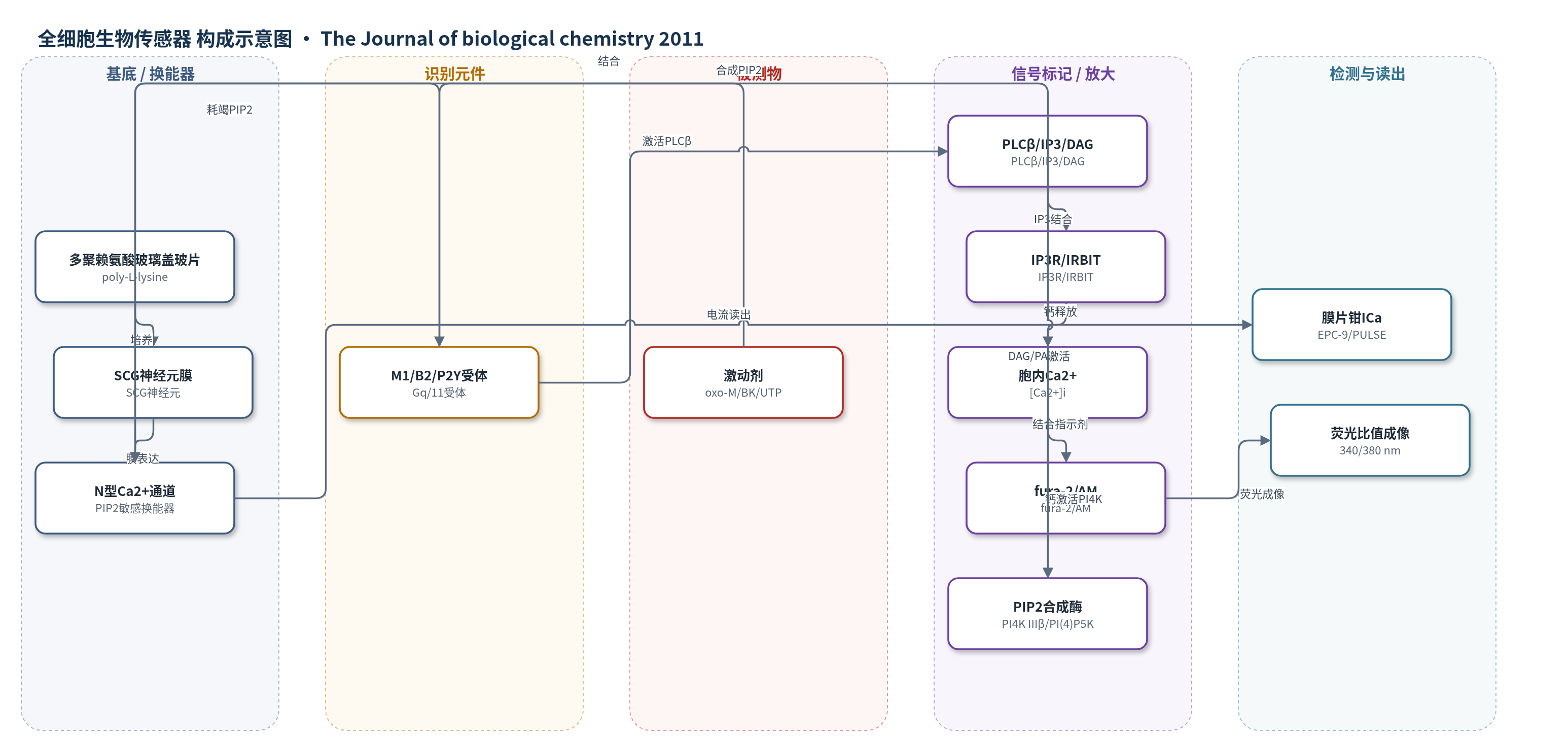
点击图片查看大图 · 依据论文自动绘制
传感器类型
全细胞生物传感器
检测对象
膜磷脂酰肌醇4,5-二磷酸(PIP2)水平、胞内钙离子([Ca2+]i);样品基质:培养的上颈神经节(SCG)交感神经元
检测原理
在SCG交感神经元中,M1、B2、P2Y受体被oxo-M、BK、UTP激活后,PLCβ水解PIP2生成IP3和DAG。IP3在IP3R附近达到阈值时克服IRBIT抑制,释放内质网Ca2+,fura-2/AM的340/380 nm比值升高。Ca2+经NCS-1激活PI 4-激酶IIIβ,DAG经DAG激酶生成PA,并与Rho/Rho激酶协同激活PI(4)P 5-激酶,促进PIP2合成。N型Ca2+通道结合PIP2,ICa随PIP2水平变化:M1/AT1远离IP3R,局部IP3低,PIP2耗竭、ICa受抑;B2/P2Y与IP3R共定位,Ca2+触发PIP2补偿合成,ICa维持。
检测灵敏度
—
效应效果
对照神经元中,oxo-M使ICa抑制57±3%,而UTP和BK仅3.3±0.5%和5.5±0.8%;阻断PI 4-激酶IIIβ、RhoA、Rho激酶或DAG激酶后,UTP/BK对ICa抑制升至23–43%。fura-2成像显示UTP/BK引起340/380 nm比值升高0.12±0.01和0.25±0.02,oxo-M仅0.02±0.01;过表达M1后oxo-M升至0.17±0.02。WT IRBIT使UTP/BK应答细胞比例由86%降至67%/58%,IRBIT-S68A使oxo-M应答比例由15%升至64%、幅度由0.02升至0.09。结果支持通道/荧光双读出具有受体选择性,但未报告实际样品回收率或ELISA对比。
传感器的构成
- 基底/培养载体:多聚赖氨酸(poly-L-lysine)包被玻璃盖玻片,用于培养上颈神经节(SCG)交感神经元。
- 换能器/识别元件:N型电压门控Ca2+通道(N-type CaV channels),直接结合膜PIP2,其电流ICa随PIP2水平变化,作为PIP2生物传感器。
- 刺激/识别元件:Gq/11偶联受体M1、B2、P2Y(及AT1),结合激动剂oxo-M、BK、UTP,启动PLCβ信号。
- 信号标记物:fura-2/AM荧光Ca2+指示剂,用于340/380 nm比值成像检测[Ca2+]i。
- 转染标记:EGFP绿色荧光蛋白,用于标记转染神经元并作为对照。
- 调控/抑制元件:PIK93、Y27632、DAG-kinase inhibitor II、DN PI 4-kinase IIIβ D656A、DN RhoA T19N、WT/DN IRBIT,用于阻断PIP2合成或IP3R阈值。
- 读出装置:穿孔膜片钳(amphotericin B)配合EPC-9放大器/PULSE软件记录ICa;倒置荧光显微镜/CCD记录fura-2比值。
中文摘要
磷脂酰肌醇4,5-二磷酸(PIP2)调节上颈神经节(SCG)交感神经元中的N型Ca2+电流(ICa)和M型K+电流。M1毒蕈碱和AT1血管紧张素受体不引起胞内Ca2+升高,并通过耗竭PIP2抑制两种电流;而B2缓激肽和P2Y嘌呤受体引起强IP3介导的[Ca2+]i升高,不耗竭PIP2也不抑制ICa。作者提出这种受体特异性源于差异化的Ca2+i信号及其对PI 4-激酶介导PIP2合成的刺激。本研究用ICa作为PIP2生物传感器,检测PI 4-激酶IIIβ、PI(4)P 5-激酶、DAG激酶、Rho/Rho激酶及IRBIT的作用。结果显示,阻断PI 4-激酶IIIβ、DAG激酶、Rho或Rho激酶后,三种受体激动剂均抑制ICa;过表达M1受体使毒蕈碱激动剂引起大Ca2+i信号;野生型IRBIT降低Ca2+i信号,显性负性IRBIT-S68A显著增强毒蕈碱反应。结论:在微区组织之上,IRBIT为IP3设置阈值,维持受体特异性。
英文摘要
Phosphatidylinositol 4,5-bisphosphate (PIP(2)) regulates Ca(2+) (I(Ca)) and M-type K(+) currents in superior cervical ganglion sympathetic neurons. In those cells, M(1) muscarinic and AT(1) angiotensin types do not elicit Ca(2+)(i) signals and suppress both currents via depletion of PIP(2), whereas the B(2) bradykinin and P2Y purinergic types elicit robust IP(3)-mediated [Ca(2+)](i) rises and neither deplete PIP(2) nor inhibit I(Ca). We have suggested that this specificity arises from differential Ca(2+)(i) signals underlying receptor-specific stimulation of PIP(2) synthesis by phosphatidylinositol (PI) 4-kinase. Here, we investigate which PI 4-kinase isoform underlies this signal, whether stimulation of PI 4-phosphate 5-kinase is also required, and the origin of receptor-specific Ca(2+)(i) signals. Recordings of I(Ca) were used as a PIP(2) "biosensor." In control, stimulation of M(1), but not B(2) or P2Y, receptors robustly suppressed I(Ca). However, when PI 4-kinase IIIβ, diacylglycerol kinase, Rho, or Rho-kinase was blocked, agonists of all three receptors robustly suppressed I(Ca). Overexpression of exogenous M(1) receptors yielded large [Ca(2+)](i) rises by muscarinic agonist, and transfection of wild-type IRBIT decreased Ca(2+)(i) signals, whereas dominant negative IRBIT-S68A had little effect on B(2) or P2Y responses but greatly increased muscarinic responses. We conclude that overlaid on microdomain organization is IRBIT, setting a "threshold" for [IP(3)], assisting in fidelity of receptor specificity.