组成图示
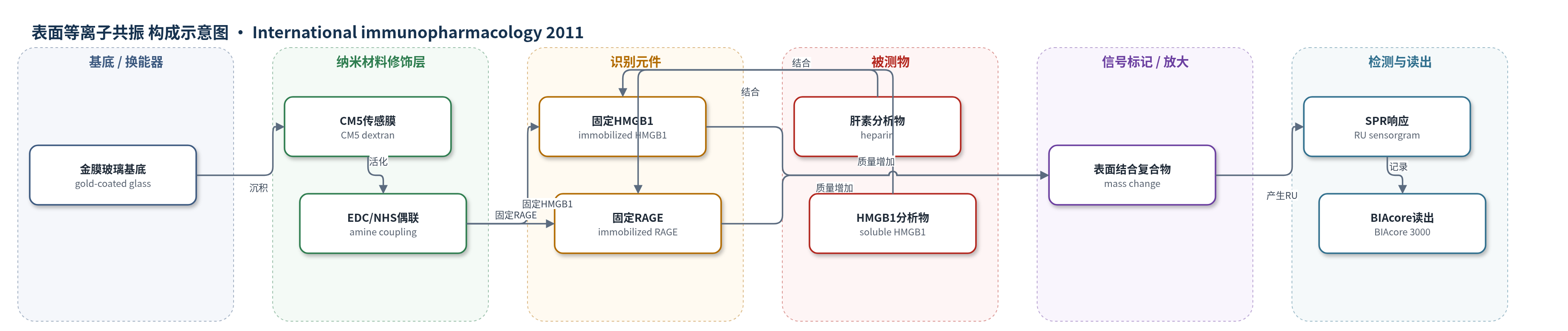
点击图片查看大图 · 依据论文自动绘制
传感器类型
表面等离子共振(SPR)生物传感器
检测对象
肝素(heparin)、高迁移率族蛋白1(HMGB1);样品基质:HBS-EP缓冲液(10 mM HEPES、150 mM NaCl、3 mM EDTA、0.005% P20,pH 7.4)。ELISA部分检测TNF-α、IL-6,样品基质为RAW264.7/HUVEC细胞上清。
检测原理
SPR生物传感器以金膜玻璃为基底,CM5羧甲基葡聚糖层通过EDC/NHS胺偶联固定HMGB1或RAGE。当肝素或HMGB1分析物流经传感表面时,与固定配体发生特异性结合,使界面质量/折射率增加,引起表面等离子共振波长偏移,被BIAcore 3000记录为响应单位(RU)随时间变化的传感图。结合相与解离相按1:1 Langmuir模型拟合,得到ka、kd和KD。肝素与HMGB1结合后改变HMGB1二级结构,使RAGE结合位点暴露或构象改变,从而降低HMGB1与RAGE的结合量,表现为混合肝素后RU下降。该方法无需外源标记,信号直接反映结合事件。
检测灵敏度
原文未报告LOD、线性范围、灵敏度斜率、相关系数。
效应效果
SPR固定量显示HMGB1约9000 RU(约9 ng/mm2),RAGE约5000 RU(约5 ng/mm2)。肝素与RAGE无结合,10000 U/L时响应低于10 RU,说明选择性较好。肝素-HMGB1结合KD=4.5×10⁻⁹ mol/L,HMGB1-RAGE结合KD=9.77×10−8 mol/L。加入肝素后HMGB1/RAGE结合RU由约100 RU降至约50 RU,不同肝素浓度对应ΔRU为55、62、50、48和2 RU。ELISA显示肝素抑制RAW264.7和HUVEC中TNF-α、IL-6释放,10 U/L和50 U/L抑制最明显,50 U/L在HUVEC中约抑制HMGB1效应的50%(P<0.05)。作者认为低剂量肝素可通过改变HMGB1构象发挥抗炎作用。
传感器的构成
- 基底/换能器:金膜玻璃表面(gold-coated glass surface),支持SPR并产生光学信号。
- 传感膜:CM5羧甲基葡聚糖层(carboxymethylated dextran),提供羧基用于蛋白固定。
- 偶联试剂:EDC/NHS胺偶联试剂盒(EDC/NHS amine-coupling kit),活化羧基并共价连接蛋白氨基。
- 识别元件A:固定HMGB1(immobilized HMGB1,Fc2),作为肝素结合配体。
- 识别元件B:固定RAGE(immobilized RAGE,Fc4),作为HMGB1结合配体。
- 运行缓冲液:HBS-EP(10 mM HEPES、150 mM NaCl、3 mM EDTA、0.005% P20,pH 7.4),维持结合条件并输送样品。
- 再生液:10 mM NaOH,解离结合物并恢复传感表面。
- 读出系统:BIAcore 3000 SPR系统,记录响应单位(RU)传感图。
中文摘要
高迁移率族蛋白1(HMGB1)是炎症晚期介质,其细胞因子活性主要由晚期糖基化终末产物受体(RAGE)介导。由于HMGB1具有肝素结合活性,本研究探讨肝素是否干扰HMGB1/RAGE相互作用并抑制其细胞因子活性。作者采用荧光光谱、圆二色谱和表面等离子共振(SPR)生物传感器技术评估肝素对HMGB1构象及HMGB1/RAGE结合的影响。不同浓度肝素(0、50、100和1000 U/L)处理后,HMGB1荧光峰强度升高且发射波长红移;圆二色谱显示β-折叠含量下降、α-螺旋含量增加,提示构象改变。SPR测定肝素与HMGB1的平衡解离常数KD为4.5×10−9 mol/L,HMGB1与RAGE的KD为9.77×10−8 mol/L,肝素与RAGE无结合。肝素处理后HMGB1与RAGE的结合量下降。ELISA显示肝素抑制RAW264.7巨噬细胞和人脐静脉内皮细胞(HUVEC)中TNF-α和IL-6释放,10 U/L和50 U/L分别抑制最明显。结论:肝素通过改变HMGB1构象降低HMGB1/RAGE亲和力,低剂量肝素即可发挥较好抗炎效果。
英文摘要
High-mobility group protein 1 (HMGB1) has been identified as a late-acting mediator of inflammation. The receptor for advanced glycation end products (RAGE) is the main receptor and mediates the cytokine activity of HMGB1. Since HMGB1 also exhibits heparin-binding activity, we investigated whether heparin interferes with HMGB1/RAGE interaction and prevents the cytokine activity. We used fluorescence spectrometry, circular dichroism spectrometry and SPR biosensor technique to evaluate the effect. After treatment of HMGB1 with different concentrations of heparin (0, 50, 100 and 1000 U/L), the fluorescence peak values of HMGB1 increased and the emission wavelength showed red shifts; further, the secondary structure of HMGB1 showed a marked change in that the content of β-pleated sheet reduced while that of α-helix increased. The equilibrium dissociation constants (K(D)) were determined by SPR technique; K(D)=4.5 × 10(-9)mol/L for heparin and HMGB1 and K(D)=9.77 × 10(-8)mol/L for HMGB1 and RAGE, respectively. Heparin and RAGE had no interaction. The amount of HMGB1 and RAGE bound forms reduced after treatment with heparin. ELISA revealed that addition of heparin inhibited the TNF-α and IL-6 released by macrophages RAW264.7 and HUVEC; 10 U/L and 50 U/L of heparin showed the most marked inhibitory effect in RAW264.7 cells and in HUVEC, respectively. In conclusion, heparin can combine with HMGB1 and affect the affinity of HMGB1/RAGE by changing the conformation of HMGB1. And this effect was independent of heparin concentration, so that a low dose of heparin was sufficient to achieve the best anti-inflammatory effect in our test.