组成图示
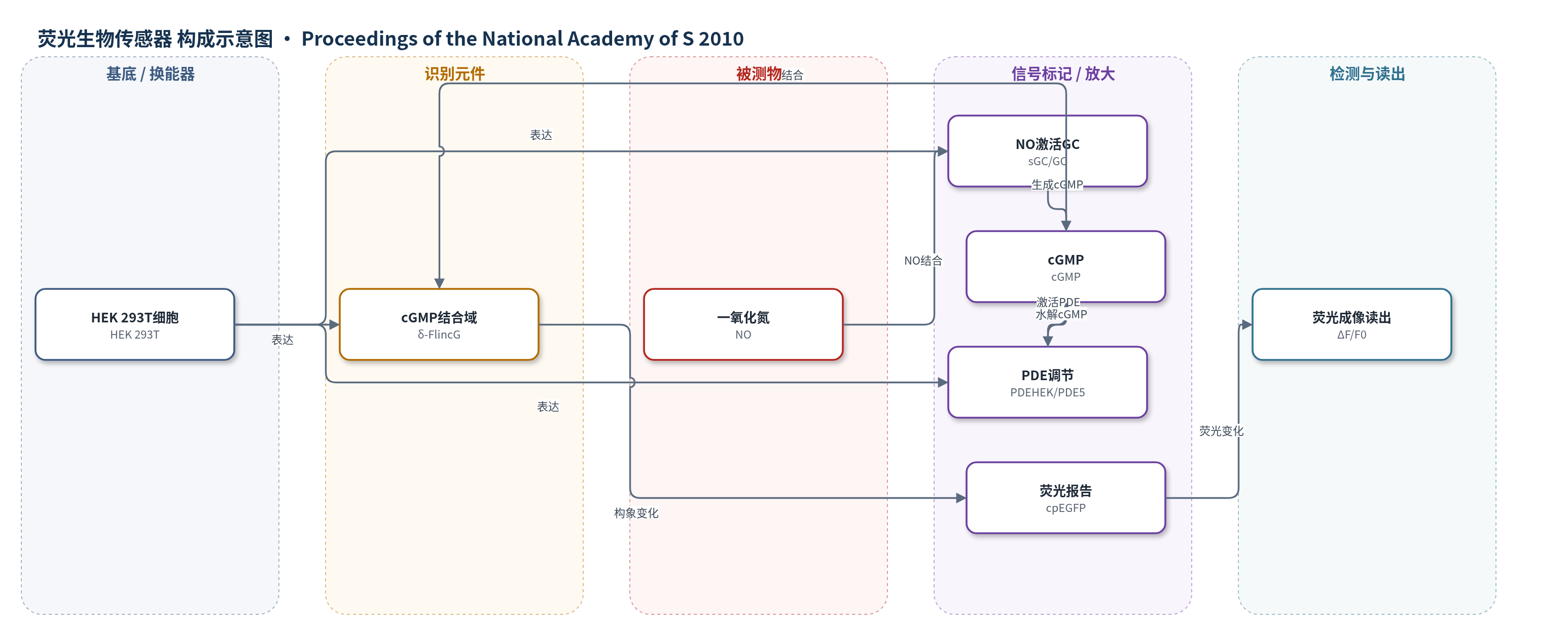
点击图片查看大图 · 依据论文自动绘制
传感器类型
荧光生物传感器
检测对象
一氧化氮(NO)、环磷酸鸟苷(cGMP);样品基质:HEK 293T 细胞培养液/细胞内环境
检测原理
NO 扩散进入 HEK 细胞后,与 NO 激活鸟苷酸环化酶(sGC)的血红素结合位点结合,使 GC 构象激活并催化 GTP 生成 cGMP。cGMP 浓度升高后,结合 δ-FlincG 中来自 cGMP 依赖性蛋白激酶的双 cGMP 结合域,引起融合 cpEGFP 的构象/荧光变化,经显微镜以 ΔF/F0 读出。PDEHEK 或 PDE5 水解 cGMP,使信号衰减;PDE5 被 cGMP 慢速激活并形成持续活性磷酸化形式,产生负反馈。由于细胞内 sGC 受体过量、酶联受体在极低 NO 下具有高转导效率,以及 GC/PDE 活性比例不同,信号被放大,因此 pM 级 NO 即可产生可测荧光响应。
检测灵敏度
可检测 NO 低至 1 pM;共表达 PDE5 细胞可检测 10 pM;100 ms 喷注计算峰值胞内 NO 23 pM 可检测;3 pM NO 产生约 0.13 μM cGMP 信号。
效应效果
δ-FlincG 对 cGMP 相对 cAMP 选择性高,动力学快,可实时定量 cGMP。NO 响应被 ODQ 抑制,IBMX 主要减慢衰减,说明上升相反映 GC 活性。GClowPDEHEK 细胞 NO 上升相 EC50 为 6.9 nM,模型预测 6.0 nM;30 nM NO 后脱敏约 37±1%,模型 38%。去除 CPTIO 检测到环境 NO 约 12 pM。PDE5 细胞可检测 10 pM NO,PDE5 恢复半时约 10 min,10 min 后恢复不完全。模型给出 GChighPDE5low 与血小板放大倍数约 20,000,小脑星形胶质细胞约 0.3×10^6,支持生理 pM 级 NO 研究。
传感器的构成
- 细胞基底:HEK 293T 细胞(HEK),作为传感器表达与 NO-cGMP 信号转导的细胞环境
- 识别元件:δ-FlincG 中 cGMP 依赖性蛋白激酶双 cGMP 结合域,特异性结合 cGMP
- 荧光换能器:循环置换 EGFP(cpEGFP),cGMP 结合引起荧光变化
- 信号生成元件:NO 激活鸟苷酸环化酶(sGC/GC),NO 结合后催化 cGMP 生成
- 信号调节元件:磷酸二酯酶(PDEHEK/PDE5),水解 cGMP 并负反馈调节信号
- 荧光读出:倒置显微镜与相机,记录 ΔF/F0 荧光变化
中文摘要
一氧化氮(NO)是体内广泛存在的可扩散神经递质,通过结合含鸟苷酸环化酶(GC)转导域的受体,使靶细胞中 cGMP 积累。尽管该通路重要,但其在生理 NO 浓度下的实时机制仍不清楚。本研究利用新型 cGMP 荧光生物传感器 δ-FlincG,将其表达于含有不同水平 NO 激活 GC 和 cGMP 水解磷酸二酯酶(PDE)活性的细胞中,系统评估其对 NO 的响应。结果显示,当 cGMP 升至具有生理意义的 30 nM 以上时,细胞对低至 1 pM 的 NO 即产生响应,使其成为迄今最灵敏的 NO 检测细胞体系;即使共表达 cGMP 激活型 PDE5 的细胞,也能响应低至 10 pM 的 NO。通过局部微管喷注短时 NO,揭示了 NO 捕获与信号转导动力学:仅持续 100 ms、计算峰值胞内 NO 浓度约 23 pM 的喷注即可被检测。上述结果可纳入细胞 NO-cGMP 信号定量模型,该模型复现了传统粗 cGMP 测量结果,并解释了 NO 在极低浓度下仅占据极少量受体即可产生生理效应的原因。
英文摘要
Nitric oxide (NO) functions as a diffusible transmitter in most tissues of the body and exerts its effects by binding to receptors harboring a guanylyl cyclase transduction domain, resulting in cGMP accumulation in target cells. Despite its widespread importance, very little is known about how this signaling pathway operates at physiological NO concentrations and in real time. To address these deficiencies, we have exploited the properties of a novel cGMP biosensor, named δ-FlincG, expressed in cells containing varying mixtures of NO-activated guanylyl cyclase and cGMP-hydrolyzing phosphodiesterase activity. Responsiveness to NO, signifying a physiologically relevant rise in cGMP to 30 nM or more, was seen at concentrations as low as 1 pM, making cells by far the most sensitive NO detectors yet encountered. Even cells coexpressing phosphodiesterase-5, a cGMP-activated isoform found in many NO target cells, responded to NO in concentrations as low as 10 pM. The dynamics of NO capture and signal transduction was revealed by administering timed puffs of NO from a local pipette. A puff lasting only 100 ms, giving a calculated peak intracellular NO concentration of 23 pM, was detectable. The results could be encapsulated in a quantitative model of cellular NO-cGMP signaling, which recapitulates the NO responsiveness reported previously from crude cGMP measurements on native cells, and which explains how NO is able to exert physiological effects at extremely low concentrations, when only a tiny proportion of its receptors would be occupied.