组成图示
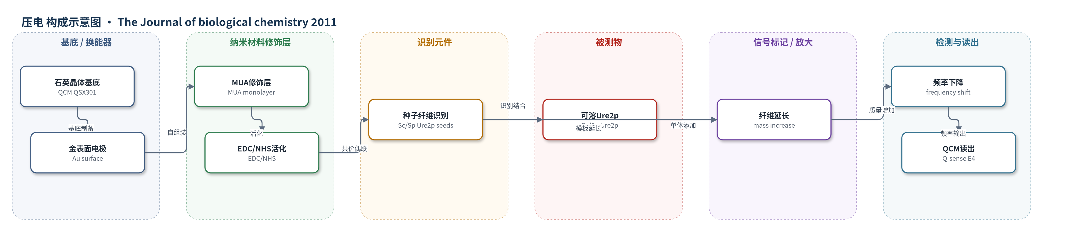
点击图片查看大图 · 依据论文自动绘制
传感器类型
压电(QCM)生物传感器
检测对象
可溶 Ure2p 蛋白(ScUre2p、SpUre2p);样品基质:50 mM Tris-HCl pH 8.4、200 mM NaCl 缓冲液
检测原理
该研究将 ScUre2p 或 SpUre2p 种子纤维经 MUA/EDC-NHS 化学共价固定于 QCM 金表面,并用乙醇胺封闭。当含可溶 Ure2p 的缓冲液流经传感器时,可溶单体识别并结合到表面固定种子纤维末端,沿纤维模板发生延长。每添加一个蛋白单体,表面质量增加,使压电石英晶体共振频率下降;频率随时间线性下降的斜率反映纤维延长速率。溶液 ThT 荧光则与纤维蛋白量呈线性关系,用于整体聚合动力学。通过交叉种子实验和主方程拟合,可分离延长与断裂速率,从而比较不同 Ure2p 同源物的朊病毒传播相关参数。
检测灵敏度
未报告 LOD、线性范围、灵敏度斜率或相关系数。
效应效果
QCM 直接监测到 ScUre2p 与 SpUre2p 纤维延长时频率线性下降,AFM 证实质量增加来自纤维长度增加,而非非特异吸附。QCM 趋势与 ThT 溶液交叉种子实验一致,验证了溶液分析可分离延长和断裂步骤。比率方法在独立重复中高度可重复,但绝对参数因种子末端数差异约 5 倍波动。结果显示 SpUre2p 延长速率高于 ScUre2p,而 ScUre2p 断裂速率更高,差异约 2 倍;SpUre2p 因断裂少导致种子增殖慢,朊病毒传播能力降低。该工作为理解淀粉样纤维可传播性与不可传播性提供定量框架。
传感器的构成
- 换能器基底:石英晶体微天平(QCM, Q-sense QSX 301)金表面,提供压电换能与质量传感
- 自组装修饰层:巯基十一烷酸(MUA)在乙醇中形成有序单分子层,提供羧基
- 活化层:EDC/NHS 活化羧基,用于与种子纤维氨基共价偶联
- 识别/模板元件:ScUre2p 或 SpUre2p 种子纤维(0.5 μM,30 min 偶联),作为纤维延长模板
- 封闭层:乙醇胺(500 mM,pH 10.0,1 h)封闭未反应基团,减少非特异吸附
- 样品/被测物:可溶 ScUre2p 或 SpUre2p(20 μM)在 50 mM Tris-HCl pH 8.4、200 mM NaCl 中流经表面
- 信号读出:QCM 共振频率及三次谐波(n=3)监测质量增加
中文摘要
肽和蛋白具有自组装成淀粉样纤维的固有倾向,某些纤维可在生物体内作为朊病毒自我传播,并向宿主传递特征。某些淀粉样物种能作为朊病毒而另一些不能的机制尚未完全理解。酵母朊病毒系统实验上高度可操作,但组装过程的生物物理与动力学定量理解仍具挑战。本文研究酿酒酵母(S. cerevisiae)和 paradoxus 酵母(S. paradoxus)Ure2p 的两个近缘同源物,结合动力学理论与溶液及生物传感器检测,比较朊病毒纤维组装各微观步骤的速率。发现对这些蛋白,断裂速率编码于种子纤维结构中,而延长速率主要由可溶前体蛋白性质决定。结果进一步显示,延长更快但断裂更少的纤维可能失去作为朊病毒传播的能力。这些发现阐明体外蛋白聚集与体内朊病毒增殖之间的联系,并为定量理解正常和异常生物学途径中淀粉样纤维行为参数提供框架。
英文摘要
Peptides and proteins possess an inherent propensity to self-assemble into generic fibrillar nanostructures known as amyloid fibrils, some of which are involved in medical conditions such as Alzheimer disease. In certain cases, such structures can self-propagate in living systems as prions and transmit characteristic traits to the host organism. The mechanisms that allow certain amyloid species but not others to function as prions are not fully understood. Much progress in understanding the prion phenomenon has been achieved through the study of prions in yeast as this system has proved to be experimentally highly tractable; but quantitative understanding of the biophysics and kinetics of the assembly process has remained challenging. Here, we explore the assembly of two closely related homologues of the Ure2p protein from Saccharomyces cerevisiae and Saccharomyces paradoxus, and by using a combination of kinetic theory with solution and biosensor assays, we are able to compare the rates of the individual microscopic steps of prion fibril assembly. We find that for these proteins the fragmentation rate is encoded in the structure of the seed fibrils, whereas the elongation rate is principally determined by the nature of the soluble precursor protein. Our results further reveal that fibrils that elongate faster but fracture less frequently can lose their ability to propagate as prions. These findings illuminate the connections between the in vitro aggregation of proteins and the in vivo proliferation of prions, and provide a framework for the quantitative understanding of the parameters governing the behavior of amyloid fibrils in normal and aberrant biological pathways.