组成图示
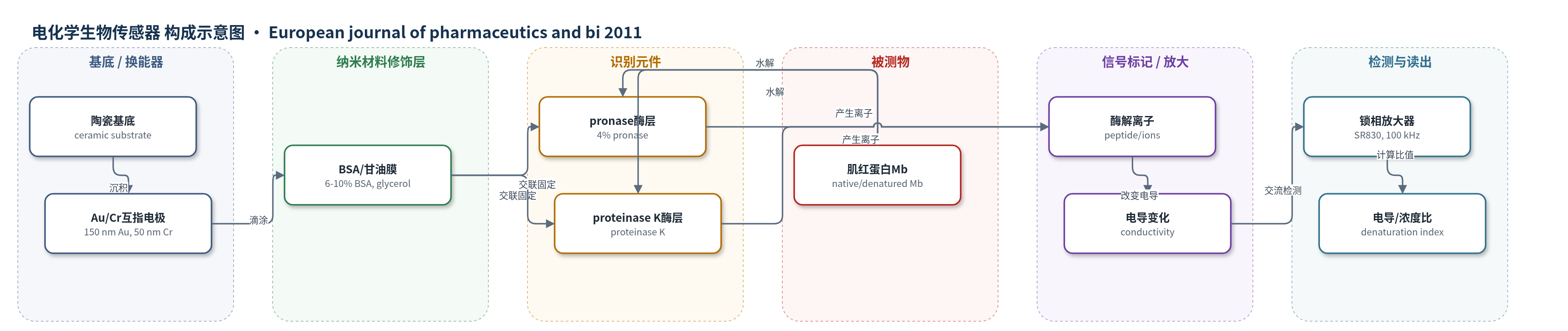
点击图片查看大图 · 依据论文自动绘制
传感器类型
电化学生物传感器
检测对象
肌红蛋白(myoglobin, Mb,天然/变性构象);样品基质:PBS释放液/蛋白溶液(PBS pH 7.4释放介质,测量缓冲液pH 7.2)
检测原理
工作电极上以戊二醛交联固定枯草溶菌素(pronase)和蛋白酶K(proteinase K)双酶膜。肌红蛋白(Mb)扩散进入酶膜后,被两种蛋白酶协同水解,产生带电荷肽段和小分子离子;离子在酶膜与缓冲介质中迁移,改变金互指电极间的电导。仪器施加10 mV、100 kHz交流电压,差分测量工作电极与参比电极的电导变化。天然Mb呈紧密球状,酶切位点可及性较低,单位浓度电导响应较小;变性Mb结构展开,更多酶切位点暴露,水解产生更多离子,电导/浓度比升高。结合Bradford或UV/Vis定量Mb浓度,通过电导/浓度比判断蛋白完整性与变性程度。
检测灵敏度
LOD: 0.3 mg·L−1;线性范围: 0–7 mg·L−1;灵敏度: 0.6913 μS·mg−1·L;R² = 0.9974;热变性Mb斜率: 2.55 μS·mg−1·L;溶剂变性Mb斜率: 3.52 μS·mg−1·L;R² = 0.9974
效应效果
该双酶电导生物传感器在0–7 mg·L−1范围内线性良好,R²=0.9974,LOD为0.3 mg·L−1;稳态响应时间约5 min,10 min内达到平衡。同一传感器同一天5次测量的变异系数为5.5%–6.4%,4 ℃保存至少1个月仍稳定。差分参比电极可抑制非特异响应。与UV/Vis吸光度比值法相比,两种方法对MI–MIV微球释放Mb的构象判断一致;UV/Vis可定位血红素或珠蛋白部分变化,但不能准确定量变性蛋白,而生物传感器结合Bradford定量可给出电导/浓度比,反映变性程度。作者认为该方法快速、适用于不同大小蛋白(如BSA 67 kDa),但难以检测可溶性聚集体,需配合SEC或SDS–PAGE。
传感器的构成
- 基底/换能器电极:陶瓷基底(ceramic substrate)上真空沉积50 nm Cr粘附层与150 nm Au互指薄膜电极,作为电导换能器
- 识别/催化层1:PBS中4% pronase、6% BSA、10%甘油滴涂于工作电极,经GA蒸气交联,提供蛋白酶水解位点
- 识别/催化层2:PBS中proteinase K、6% BSA、10%甘油滴涂于工作电极,经GA蒸气交联,与pronase协同水解Mb
- 参比封闭层:10% BSA与10%甘油滴涂于参比电极并GA交联,用于差分测量与非特异响应抑制
- 交联/稳定剂:戊二醛(GA)蒸气交联酶膜;BSA与甘油稳定酶并维持膜结构
- 测量介质:5 mM PBS(pH 7.2)提供离子缓冲环境,使酶解产生的离子改变电导
中文摘要
本研究采用多重乳化-溶剂萃取/蒸发法制备负载肌红蛋白(Mb)的聚己内酯(PCL)微球,并在不同包封工艺条件下考察蛋白完整性。作者同时使用特定波长紫外-可见吸光度比值法和基于蛋白酶K与枯草溶菌素(pronase)的电导型双酶生物传感器对释放的Mb构象进行监测。结果表明,在标准工艺条件下,Mb基本保持天然构象;当改变包封条件时,可观察到不同程度的蛋白变性。其中,在减压条件下快速去除溶剂以及加入较高分子量PCL均会导致蛋白结构改变。对于减压除溶剂体系,蛋白完整性损失可归因于颗粒固化过快而将残留有机溶剂包埋于微球内部;对于加入高分子量PCL的体系,变性可解释为有机相黏度升高使蛋白在水/有机溶剂界面暴露时间延长。该工作说明两种分析方法均可用于快速评估可降解微球中蛋白的完整性。
英文摘要
Myoglobin (Mb)-loaded poly(ε-caprolactone) (PCL) microparticles were prepared by multiple emulsion with solvent extraction/evaporation method under more or less deleterious operating conditions. The protein integrity was monitored using both UV/Vis absorbance ratio method at specific wavelengths and a conductometric bi-enzymatic biosensor based on proteinase K and pronase. Under standard operating conditions, Mb remained in native conformation, while different degrees of protein denaturation were observed by changing the encapsulation conditions. It was shown that solvent elimination under reduced pressure and in a lower extent addition of a higher molecular weight PCL led to protein alteration. In the first case, the loss of protein integrity can be attributed to residual solvent entrapped in particles whose solidification was accelerated. In the second case, denaturation may be explained by an increase in the protein exposure time at water/organic solvent interface due to an increase in organic phase viscosity.