组成图示
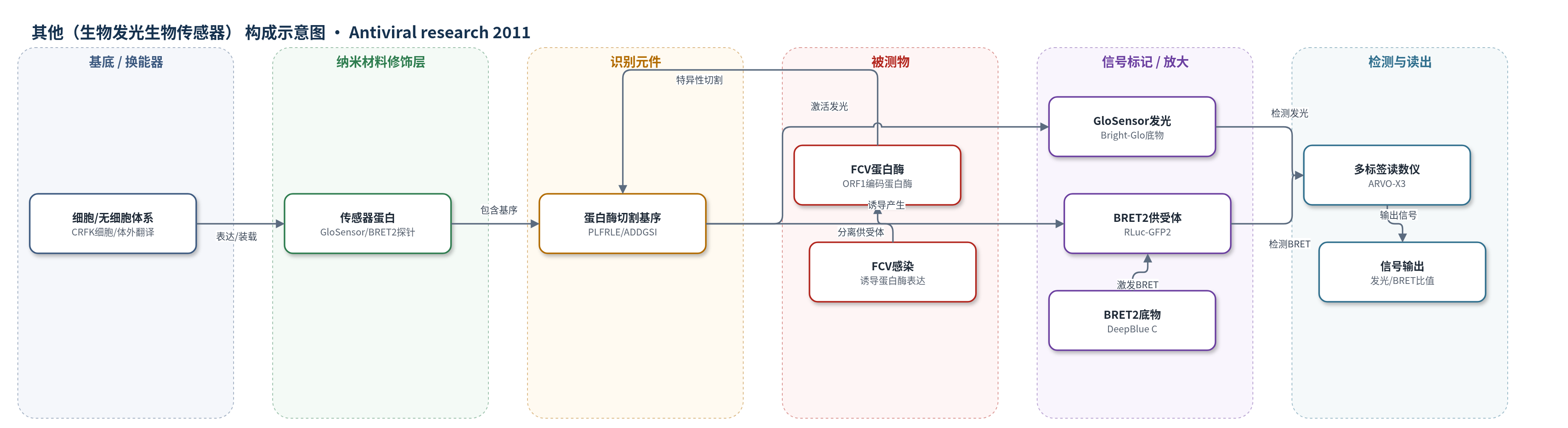
点击图片查看大图 · 依据论文自动绘制
传感器类型
其他(生物发光生物传感器)
检测对象
猫杯状病毒蛋白酶(feline calicivirus protease, FCV protease)活性、猫杯状病毒感染(feline calicivirus infection, FCV);样品基质:无细胞体外翻译体系、CRFK 猫肾细胞培养物/感染细胞。
检测原理
该传感器以 FCV 蛋白酶切割基序 PLFRLE/ADDGSI 为识别元件。GloSensor 体系中,FCV 蛋白酶识别并切割重排萤火虫荧光素酶中的该基序,解除构象约束,使萤火虫荧光素酶活性增强;加入 Bright-Glo 底物后产生生物发光,发光强度随蛋白酶量或 FCV 感染滴度升高而增加。BRET2 体系中,RLuc 供体与 GFP2 受体邻近,加入 DeepBlue C 后 RLuc 发光能量转移至 GFP2,产生 515 nm 荧光;蛋白酶切割中间基序使供受体分离,BRET2 比值(GFP2/RLuc)下降,下降幅度随 FCV 滴度升高而增大。突变基序不能被切割,因此无信号变化,保证检测特异性。
检测灵敏度
原文未报告 LOD、线性范围、灵敏度斜率或相关系数。
效应效果
特异性方面,突变切割基序传感器和失活 FCV 蛋白酶均无信号变化,表明信号依赖 FCV 蛋白酶活性。无细胞 GloSensor 在 1:4 蛋白酶稀释下最大发光比达 193 倍。细胞体系中,CellGlo 在 MOI 0.1、0.01、0.001 时发光比分别为 37.9、38.3、10.3 倍;BRET2 在 MOI 1、0.1、0.01 时 BRET 比值分别为 0.22、0.48、0.82,CellGlo 灵敏度至少高 10 倍。利巴韦林在 CellGlo 细胞中 50、100、200、400 μM 抑制率为 37.2%、82.3%、97.9%、99.9%,IC50 为 61 μM;BRET2 细胞中为 27.3%、61.4%、90.9%、100%,IC50 为 79 μM。400 μM 细胞毒性可忽略。作者认为可用于高通量筛选抗 FCV 蛋白酶和抗病毒化合物。
传感器的构成
- 换能器/信号蛋白:GloSensor(工程化萤火虫荧光素酶,含 FCV 蛋白酶切割基序 PLFRLE/ADDGSI),切割后构象激活发光。
- 识别元件:FCV 蛋白酶切割基序 PLFRLE/ADDGSI(VP1 前体 P6–P6′ 位点),被 FCV 蛋白酶特异性切割;突变基序 PLFRLA/ADDGSI 作阴性对照。
- 信号标记/供受体:BRET2 探针中的 RLuc(Renilla luciferase)供体与 GFP2 受体,中间插入切割基序,切割后分离导致 BRET2 比值下降。
- 发光底物:Bright-Glo Luciferase Assay Substrate(GloSensor 发光底物)或 Coelenterazine 400A/DeepBlue C(BRET2 底物),提供发光或能量转移信号。
- 表达/细胞平台:CRFK 猫肾细胞稳定表达 CellGlo-FCV-Cut/Uncut 或 BRET2-FCV-Cut/Uncut;无细胞体系用小麦胚芽/兔网织红细胞体外转录翻译表达传感器和 FCV 蛋白酶。
- 检测载体:96 孔板(ViewPlate black-96/white-96),承载反应体系并适配多标签读数仪。
- 读出装置:2030 Multilabel Reader ARVO-X3,测量 GloSensor 生物发光或 BRET2 双通道发光(RLuc 410 nm、GFP2 515 nm)。
中文摘要
猫杯状病毒(FCV)是重要兽医病原体,可引起猫呼吸道疾病,并因细胞培养良好而常作为不可培养杯状病毒模型。本研究建立无细胞和两种细胞培养生物传感器体系,检测 FCV 蛋白酶活性。体系分别基于荧光素酶传感器(GloSensor)和第二代生物发光共振能量转移(BRET2)。GloSensor 在重排萤火虫荧光素酶中插入 FCV 蛋白酶切割基序;BRET2 探针在 Renilla 荧光素酶(RLuc)和绿色荧光蛋白变体(GFP2)之间插入相同基序。含突变切割基序的传感器作为阴性对照。无细胞检测中,GloSensor 发光随 FCV 蛋白酶量增加而增强,突变基序无信号变化。稳定表达传感器的猫细胞系中,GloSensor 发光随 FCV 滴度升高而增加,BRET2 信号随 FCV 滴度升高而降低,突变基序细胞无显著变化。两种传感器均可检测利巴韦林对 FCV 感染细胞的抑制作用。结果表明,这些生物传感器可用于监测感染细胞中 FCV 蛋白酶活性,并适用于无细胞体系和培养细胞中抗 FCV 化合物筛选。
英文摘要
Feline calicivirus (FCV) is an important veterinary pathogen and causes respiratory disease in cats. Because it grows well in cell culture, FCV is often used as a model virus of non-culturable caliciviruses. In this study, a cell-free and two cell culture-based biosensor assay systems were established to detect FCV protease activity. The assays utilize luciferase sensor technology or second-generation bioluminescence resonance energy transfer (BRET2). A luciferase sensor was designed to contain an FCV protease cleavage motif within the permutated luciferase (GloSensor). The BRET2-based probe contained the same cleavage motif flanked by a renilla luciferase and a variant of green fluorescent protein. To confirm the specificity of these assay systems, GloSensor or a BRET2-based probe containing a mutation in the cleavage motif was also constructed. In a cell-free assay, GloSensor showed increased luminescence in proportion to the amount of FCV protease, while no signal change was observed when the construct harboring the mutant cleavage motif was used. A feline cell line stably expressing GloSensor or the BRET2-based probe was established. Increased levels of GloSensor luminescence, and decreased levels of BRET2 signals were observed according to input FCV titers. In contrast, no significant signal change was observed in the cells stably expressing the mutant cleavage motif. GloSensor and the BRET2-based probe were capable of detecting the inhibitory activity of ribavirin in FCV-infected cells. Our results demonstrate that these biosensors are useful to detect FCV protease activity induced in infected cells, and well worth consideration for screening of anti-FCV protease compounds in cell-free system as well as anti-FCV compounds in cultured cells.