组成图示
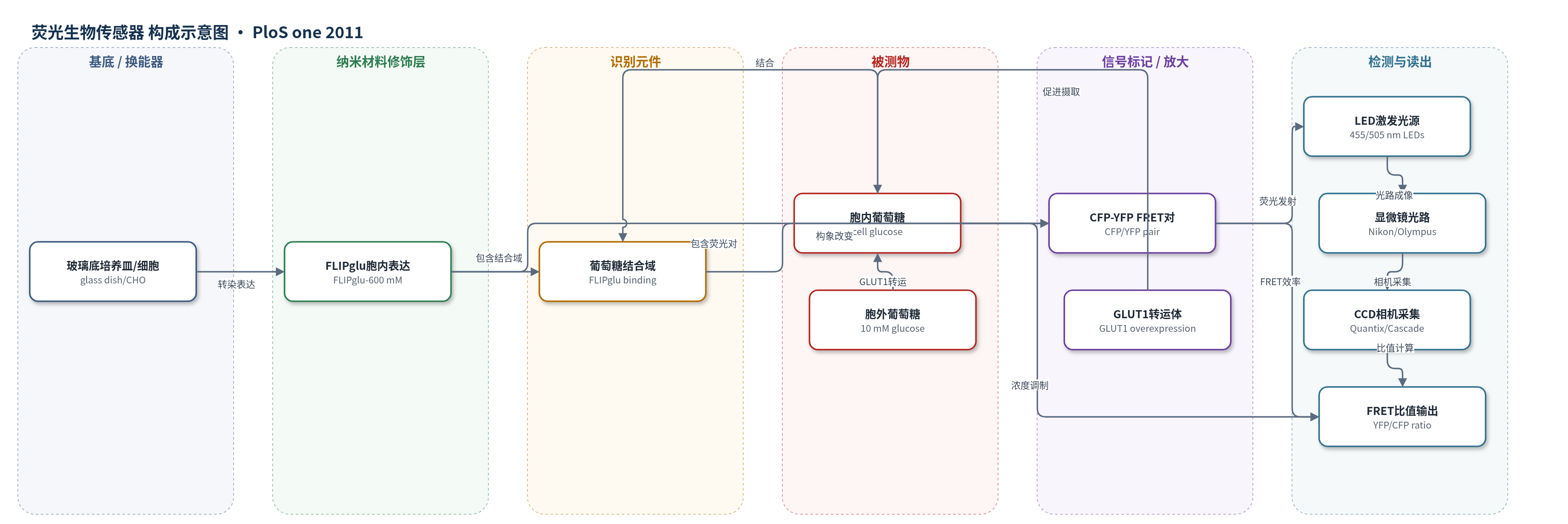
点击图片查看大图 · 依据论文自动绘制
传感器类型
荧光生物传感器
检测对象
葡萄糖(glucose);样品基质:CHO 细胞培养液/细胞内胞质(含 10 mM 葡萄糖等)
检测原理
FLIPglu-600 mM 为遗传编码 FRET 葡萄糖传感器,由 CFP 供体、YFP 受体和葡萄糖结合域组成。胞外葡萄糖经 GLUT1/GLUT4 进入 CHO 细胞后,与 FLIPglu 结合域结合,引起传感器构象变化,改变 CFP 到 YFP 的 FRET 效率;以 YFP/CFP 发射比作为读出,比值随胞内葡萄糖浓度升高而改变。过表达 GLUT1 可提高胞内葡萄糖水平,增强信号;Cyto B 阻断 GLUT 外排,使清除主要反映代谢。HKI/HKII 的亚细胞定位改变葡萄糖磷酸化与糖原合成,间接影响胞内葡萄糖清除动力学。
检测灵敏度
原文未报告 LOD、线性范围、灵敏度斜率或相关系数。
效应效果
该 FRET 葡萄糖传感器在 CHO 细胞中可实时监测胞内葡萄糖动态。过表达 GLUT1 后葡萄糖摄取时间常数由 34.5±5 s 降至 1.9±0.6 s,清除半衰期约 9.8±3.5 s;GLUT4 摄取约 2.5 s,清除 12.5±6.5 s。HKI 过表达使葡萄糖清除 t1/2 由 114±12 s 缩短至 72±7 s,HKII 则延长至 217±20 s;siRNA 敲低 HKI/HKII 均降低清除速率。FCCP 不引起 HKII 转位,提示信号依赖 G-6-P 而非 ATP;Cyto B 可阻断 GLUT 介导外排。长时间成像存在基线漂移,需指数拟合校正。作者认为该方法适用于活细胞葡萄糖代谢与 HK 定位研究。
传感器的构成
- 样品基底:玻璃底培养皿(glass bottomed Petri dish)与 CHO 细胞,承载活细胞并维持成像环境。
- 传感器表达层:CHO 细胞内表达 FLIPglu-600 mM(遗传编码 FRET 葡萄糖传感器),用于胞内葡萄糖检测。
- 识别元件:FLIPglu-600 mM 的葡萄糖结合域(原文未给出具体结合蛋白名称),结合葡萄糖后改变 FRET。
- 荧光信号元件:CFP 供体与 YFP 受体(CFP/YFP FRET 对),葡萄糖结合引起 YFP/CFP 发射比变化。
- 转运放大元件:GLUT1 或 GLUT4 过表达(GLUT1-GFP/GLUT4-GFP 构建体),增加葡萄糖摄取与胞内葡萄糖水平。
- 定位对照元件:HKI-YFP、HKII-YFP、PTG-GFP/mCherry 与 MitoTracker-YFP,用于追踪己糖激酶、糖原和线粒体。
- 读出系统:Nikon Eclipse TE300/Olympus IX70 显微镜、455/505 nm LED、CFP/YFP 滤片、Dual View 分光器、CCD/数字相机与 MetaFluor 软件,输出 YFP/CFP FRET 比值。
中文摘要
葡萄糖代谢的第一步是己糖激酶(HKs)将葡萄糖磷酸化为葡萄糖-6-磷酸(G-6-P)。HKI 主要结合线粒体,HKII 可分布于线粒体和胞质。本研究在 CHO 细胞中表达 HKI-YFP 和 HKII-YFP,同时用基于 FRET 的细胞内葡萄糖生物传感器 FLIPglu-600 mM 监测葡萄糖处理,并用 GFP 标记的糖原相关蛋白 PTG 评估糖原形成。结果显示,HKI 始终强结合线粒体;HKII 可在线粒体与胞质间随葡萄糖、G-6-P 和 Akt 信号转位,但不受 ATP 影响。代谢测量表明,HKI 主要促进糖酵解;HKII 结合线粒体时促进糖酵解,位于胞质时促进糖原合成。去除葡萄糖后糖原分解产生 G-6-P,抑制 HKII 并使其从线粒体解离。结论:胞外葡萄糖通过胞内葡萄糖、G-6-P 和 Akt 等信号动态调控 HKII 定位,从而决定葡萄糖的分解代谢或合成代谢命运;HKI 则主要承诺于糖酵解。
英文摘要
BACKGROUND: The first step in glucose metabolism is conversion of glucose to glucose 6-phosphate (G-6-P) by hexokinases (HKs), a family with 4 isoforms. The two most common isoforms, HKI and HKII, have overlapping tissue expression, but different subcellular distributions, with HKI associated mainly with mitochondria and HKII associated with both mitochondrial and cytoplasmic compartments. Here we tested the hypothesis that these different subcellular distributions are associated with different metabolic roles, with mitochondrially-bound HK's channeling G-6-P towards glycolysis (catabolic use), and cytoplasmic HKII regulating glycogen formation (anabolic use).
METHODOLOGY/PRINCIPAL FINDINGS: To study subcellular translocation of HKs in living cells, we expressed HKI and HKII linked to YFP in CHO cells. We concomitantly recorded the effects on glucose handling using the FRET based intracellular glucose biosensor, FLIPglu-600 mM, and glycogen formation using a glycogen-associated protein, PTG, tagged with GFP. Our results demonstrate that HKI remains strongly bound to mitochondria, whereas HKII translocates between mitochondria and the cytosol in response to glucose, G-6-P and Akt, but not ATP. Metabolic measurements suggest that HKI exclusively promotes glycolysis, whereas HKII has a more complex role, promoting glycolysis when bound to mitochondria and glycogen synthesis when located in the cytosol. Glycogen breakdown upon glucose removal leads to HKII inhibition and dissociation from mitochondria, probably mediated by increases in glycogen-derived G-6-P.
CONCLUSIONS/SIGNIFICANCE: These findings show that the catabolic versus anabolic fate of glucose is dynamically regulated by extracellular glucose via signaling molecules such as intracellular glucose, G-6-P and Akt through regulation and subcellular translocation of HKII. In contrast, HKI, which activity and regulation is much less sensitive to these factors, is mainly committed to glycolysis. This may be an important mechanism by which HK's allow cells to adapt to changing metabolic conditions to maintain energy balance and avoid injury.