组成图示
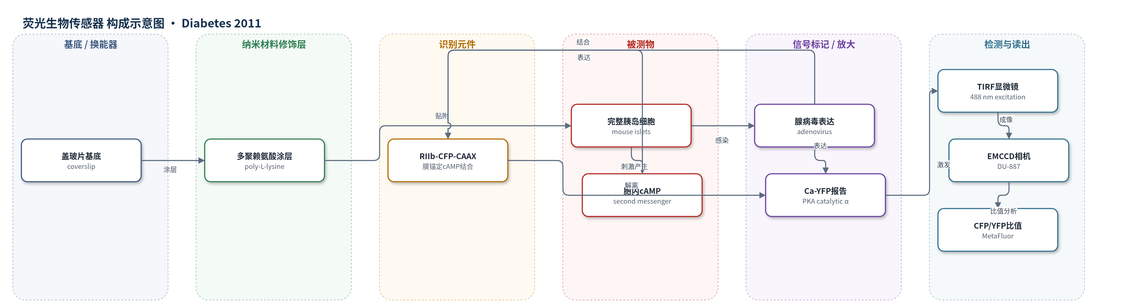
点击图片查看大图 · 依据论文自动绘制
传感器类型
荧光生物传感器
检测对象
环磷酸腺苷(cAMP);样品基质:完整小鼠胰岛(intact mouse pancreatic islets)及 MIN6 β 细胞
检测原理
腺病毒将 cAMP 转位生物传感器导入完整胰岛细胞或 MIN6 细胞。传感器由膜锚定 RIIb-CFP-CAAX 和胞质 Ca-YFP 组成:基础状态下 Ca-YFP 与 RIIb 结合,CFP/YFP 荧光比值低;当葡萄糖、胰高血糖素或 GLP-1 激活跨膜腺苷酸环化酶(tmAC)使 cAMP 升高时,cAMP 结合 RIIb,促使 Ca-YFP 从质膜附近解离,CFP 与 YFP 距离改变,CFP/YFP 比值升高。TIRF 显微镜以 488 nm 光激发近膜区域,EMCCD 相机采集 CFP 和 YFP 荧光,MetaFluor 计算背景校正比值并归一化,从而实时反映 [cAMP]pm 的升高或振荡。Ca2+ 可通过钙调蛋白敏感腺苷酸环化酶放大 cAMP 信号,但去除 Ca2+ 后振荡仍可维持,说明 cAMP 振荡并非完全依赖 Ca2+。
检测灵敏度
原文未报告 LOD、线性范围、灵敏度斜率或相关系数。
效应效果
该荧光转位生物传感器在完整小鼠胰岛中记录 α 和 β 细胞质膜下 cAMP 动力学。GLP-1 0.5–10 nmol/L 诱导 β 细胞振荡,频率 0.18–2.1 min^-1,幅度 0.06–1.5 归一化比值单位;>50 nmol/L 持续升高。胰高血糖素和 GLP-1 半最大效应约 80 和 30 nmol/L,最大效应约 1000 和 300 nmol/L。葡萄糖 11–30 mmol/L 诱导慢振荡,频率 0.05–0.4/min,幅度 0.05–0.6 比值单位。β 细胞间同步。DDA 抑制葡萄糖和激素诱导的 cAMP 升高,KH7 干扰葡萄糖氧化,CE 无持续效应;去除 Ca2+ 后振荡仍持续。应用价值:揭示完整胰岛 cAMP 信号与脉冲激素分泌调控。
传感器的构成
- 基底/支撑层:多聚赖氨酸涂层盖玻片(poly-L-lysine-coated coverslip),固定完整胰岛并作为 TIRF 成像界面
- 细胞表达层:完整小鼠胰岛(intact mouse pancreatic islets)或 MIN6 β 细胞,表达 cAMP 转位生物传感器并维持 α/β 细胞信号环境
- 识别元件:膜锚定 cAMP 结合蛋白 RIIb-CFP-CAAX(PKA 调节亚基 RIIb 融合 CFP 并 CAAX 膜锚定),特异性结合 cAMP
- 信号报告元件:PKA 催化亚基 Ca-YFP(catalytic α subunit fused to YFP),cAMP 结合后与 RIIb 解离并改变 CFP/YFP 荧光比值
- 表达载体:腺病毒(adenovirus),将 cAMP 转位生物传感器基因导入胰岛细胞或 MIN6 细胞
- 换能/读出层:TIRF 显微镜(Eclipse Ti,60× 1.45 NA)、488 nm 激发、EMCCD 相机(DU-887)与 MetaFluor 软件,输出背景校正 CFP/YFP 比值
中文摘要
cAMP 是调控胰岛 β 细胞胰岛素和 α 细胞胰高血糖素分泌的关键第二信使。分散 β 细胞可出现 cAMP 振荡,但完整胰岛内信号动力学尚不清楚。本研究利用全内反射显微镜(TIRF)和荧光转位生物传感器,记录完整小鼠胰岛外周 α 和 β 细胞质膜下 cAMP 浓度([cAMP]pm),并依据肾上腺素的相反作用鉴定细胞类型。结果显示,3 mmol/L 葡萄糖中 [cAMP]pm 低而稳定;胰高血糖素和 GLP-1 诱导剂量依赖性升高,常伴随 β 细胞间同步振荡;胰高血糖素也诱导多数 α 细胞振荡,而仅少数 α 细胞对 GLP-1 有反应。葡萄糖升至 11–30 mmol/L 可在无激素条件下诱导 α 和 β 细胞慢速 cAMP 振荡。β 细胞中 cAMP 振荡与胞质 Ca2+ 振荡协调,但并非由 Ca2+ 变化引起。跨膜腺苷酸环化酶抑制剂 DDA 抑制葡萄糖和激素诱导的 cAMP 升高,可溶性腺苷酸环化酶抑制剂 KH7 和 CE 分别干扰代谢或无效。结论:完整胰岛中受刺激 β 细胞振荡性 cAMP 信号依赖跨膜腺苷酸环化酶;α 细胞中葡萄糖和胰高血糖素诱导的 cAMP 振荡提示其参与脉冲式胰高血糖素分泌。
英文摘要
OBJECTIVE: cAMP is a critical messenger for insulin and glucagon secretion from pancreatic β- and α-cells, respectively. Dispersed β-cells show cAMP oscillations, but the signaling kinetics in cells within intact islets of Langerhans is unknown.
RESEARCH DESIGN AND METHODS: The subplasma-membrane cAMP concentration ([cAMP](pm)) was recorded in α- and β-cells in the mantle of intact mouse pancreatic islets using total internal reflection microscopy and a fluorescent translocation biosensor. Cell identification was based on the opposite effects of adrenaline on cAMP in α- and β-cells.
RESULTS: In islets exposed to 3 mmol/L glucose, [cAMP](pm) was low and stable. Glucagon and glucagon-like peptide-1(7-36)-amide (GLP-1) induced dose-dependent elevation of [cAMP](pm), often with oscillations synchronized among β-cells. Whereas glucagon also induced [cAMP](pm) oscillations in most α-cells, <20% of the α-cells responded to GLP-1. Elevation of the glucose concentration to 11-30 mmol/L in the absence of hormones induced slow [cAMP](pm) oscillations in both α- and β-cells. These cAMP oscillations were coordinated with those of the cytoplasmic Ca(2+) concentration ([Ca(2+)](i)) in the β-cells but not caused by the changes in [Ca(2+)](i). The transmembrane adenylyl cyclase (AC) inhibitor 2'5'-dideoxyadenosine suppressed the glucose- and hormone-induced [cAMP](pm) elevations, whereas the preferential inhibitors of soluble AC, KH7, and 1,3,5(10)-estratrien-2,3,17-β-triol perturbed cell metabolism and lacked effect, respectively.
CONCLUSIONS: Oscillatory [cAMP](pm) signaling in secretagogue-stimulated β-cells is maintained within intact islets and depends on transmembrane AC activity. The discovery of glucose- and glucagon-induced [cAMP](pm) oscillations in α-cells indicates the involvement of cAMP in the regulation of pulsatile glucagon secretion.