组成图示
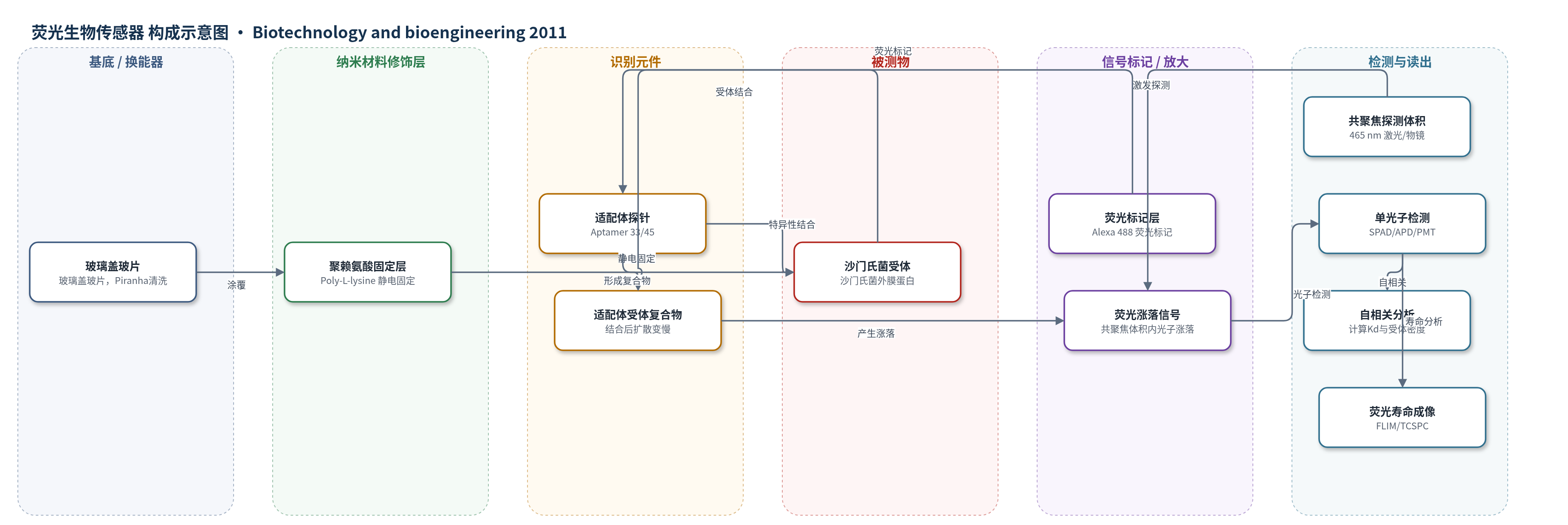
点击图片查看大图 · 依据论文自动绘制
传感器类型
荧光生物传感器
检测对象
鼠伤寒沙门氏菌(Salmonella typhimurium)外膜蛋白受体/适配体结合位点;样品基质:PBS 中活菌或聚赖氨酸固定活菌。
检测原理
荧光标记适配体在 PBS 中与活沙门氏菌表面外膜蛋白发生特异性结合。未结合适配体在共聚焦探测体积内快速扩散,结合后形成较大复合物,扩散时间变慢。FCS 通过检测荧光强度涨落并计算自相关函数,将信号分解为游离态与结合态两个扩散组分,从而获得结合分数、扩散时间和分子数。结合分数随适配体浓度变化,用单点结合方程拟合得到解离常数 Kd;在饱和浓度下,由结合分子数、共聚焦体积和细菌表面积估算受体密度。FLIM/TCSPC 利用 Alexa 488 与细菌自荧光寿命差异区分探针信号,提高单分子定量可靠性。
检测灵敏度
原文未报告 LOD、线性范围、灵敏度斜率或 R^2。报告数值:Kd = 0.1285 nM(aptamer 33)、0.3772 nM(aptamer 45);受体密度 = 42.27 ± 0.49 receptors per mm2、49.82 ± 0.45 receptors per mm2;荧光寿命:适配体结合菌 ≈ 4.015 ± 0.1170 ns,未结合菌 ≈ 3.158 ± 0.1248 ns;适配体浓度 0–5 nM,最低 0.25 nM;光束宽度 200 nm。
效应效果
该方法在活菌单细胞水平实现适配体结合定量。两种适配体对鼠伤寒沙门氏菌外膜蛋白具有特异性,并已在先前工作中对大肠杆菌外膜蛋白和脂多糖显示种属选择性。低激光功率约 8 mW 下未观察到光漂白,荧光计数稳定。Aptamer 33 与 45 的 Kd 分别为 0.1285 nM 和 0.3772 nM,受体密度分别为 42.27±0.49 和 49.82±0.45 receptors/mm2,表明表面受体数量足以支持结合。适配体结合菌荧光寿命约 4.015±0.1170 ns,未结合菌约 3.158±0.1248 ns,可用 FLIM 区分探针与自荧光。聚赖氨酸固定减少细菌移动,但长时间仍有轻微移动,导致 Kd 标准差。作者认为 FCS 可泛化用于活菌受体-配体动力学和生物传感器构建。
传感器的构成
- 基底/样品固定层:玻璃盖玻片(coverslips),经 Piranha 溶液清洗后涂覆 Poly-L-lysine,提供正电荷表面以静电固定活菌。
- 靶标/受体层:活鼠伤寒沙门氏菌(Salmonella typhimurium)细胞,其外膜蛋白(outer membrane proteins)作为适配体识别受体。
- 识别元件:Aptamer 33 与 Aptamer 45(28-mer DNA 适配体),特异性结合 S. typhimurium 外膜蛋白序列。
- 信号标记物:Alexa 488 荧光标记适配体,提供荧光信号并用于荧光寿命成像(FLIM)与 FCS 单分子分析。
- 反应介质:磷酸盐缓冲液(PBS),用于维持活菌状态和适配体结合反应。
- 光学换能/读出层:共聚焦时间分辨显微镜(Microtime 200),含 465 nm 脉冲激光、60× 水浸物镜、二向色镜、500–540 nm 发射滤片及 SPAD/APD/PMT 探测器,采集荧光涨落与寿命。
中文摘要
本文提出一种基于荧光相关光谱(FCS)的单分子实验设计,用于定量针对鼠伤寒沙门氏菌(Salmonella typhimurium)表面受体的适配体结合特性。作者利用特异性识别沙门氏菌外膜蛋白的适配体,并通过荧光标记探针,在活单细胞水平研究结合与未结合适配体的扩散动力学。通过自相关分析,可确定适配体在活细胞表面的结合动力学,并计算解离常数和受体密度。实验中,适配体被构建为结合 S. typhimurium 外膜蛋白,并评估其对大肠杆菌的特异性。荧光标记适配体探针结合 FCS 后,可在单分子灵敏度下比较结合态与游离态扩散行为,从而获得细菌受体的解离常数和受体密度。两种适配体探针的解离常数分别为 0.1285 nM 和 0.3772 nM,对应受体密度分别为 42.27 和 49.82 个/mm²。研究结果表明,表面受体数量足以支持结合,且两种适配体均具有高结合亲和力,可用于检测过程。所开发方法具有独特性,可推广用于活菌表面结合动力学和受体定量,为生物传感器构建及多种环境与生物应用中的活单细胞受体-配体动力学计算提供方法学基础。
英文摘要
This experimental design presents a single molecule approach based on fluorescence correlation spectroscopy (FCS) for the quantification of outer membrane proteins which are receptors to an aptamer specifically designed to target the surface receptors of live Salmonella typhimurium. By using correlation analysis, we also show that it is possible to determine the associated binding kinetics of these aptamers on live single cells. Aptamers are specific oligonucleotides designed to recognize conserved sequences that bind to receptors with high affinity, and therefore can be integrated into selective biosensor platforms. In our experiments, aptamers were constructed to bind to outer membrane proteins of S. typhimurium and were assessed for specificity against Escherichia coli. By fluorescently labeling aptamer probes and applying FCS, we were able to study the diffusion dynamics of bound and unbound aptamers and compare them to determine the dissociation constants and receptor densities of the bacteria for each aptamer at single molecule sensitivity. The dissociation constants for these aptamer probes calculated from autocorrelation data were 0.1285 and 0.3772 nM and the respective receptor densities were 42.27 receptors per µm(2) and 49.82 receptors per µm(2). This study provides ample evidence that the number of surface receptors is sufficient for binding and that both aptamers have a high-binding affinity and can therefore be used in detection processes. The methods developed here are unique and can be generalized to examine surface binding kinetics and receptor quantification in live bacteria at single molecule sensitivity levels. The impact of this study is broad because our approach can provide a methodology for biosensor construction and calculation of live single cell receptor-ligand kinetics in a variety of environmental and biological applications.