组成图示
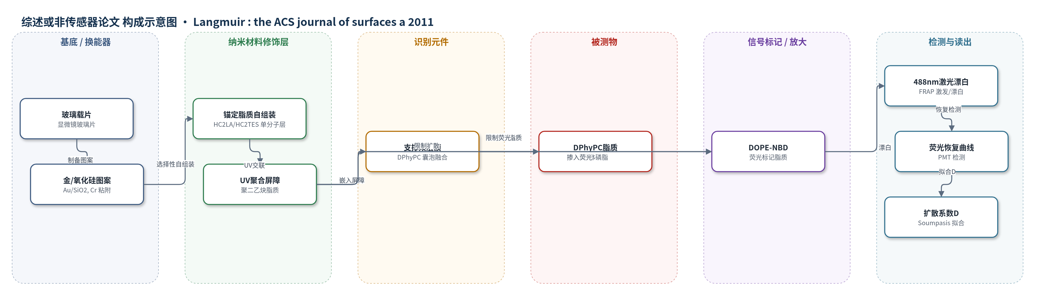
点击图片查看大图 · 依据论文自动绘制
传感器类型
综述或非传感器论文
检测对象
无明确分析物;表征对象为支撑脂质双层膜中的脂质侧向扩散(lipid lateral diffusion),样品基质为 PBS 缓冲液中的模型膜。
检测原理
该体系并非基于分析物浓度产生电信号,而是通过膜图案化与荧光漂白恢复(FRAP)表征脂质侧向扩散。首先,纳米球光刻制备金/二氧化硅超平图案基底;可聚合二乙炔基锚定脂质选择性自组装于金或氧化物区域,紫外光引发二乙炔拓扑聚合,形成聚二乙炔共轭骨架,作为不可移动扩散屏障。随后,含 DPhyPC 和 DOPE-NBD 的小单层囊泡融合到基底上,形成流体支撑双层膜。FRAP 实验中,488 nm 激光漂白局部 DOPE-NBD 荧光,周围荧光脂质扩散进入漂白区,荧光随时间恢复。恢复曲线按 Soumpasis 模型拟合得到扩散系数 D。屏障尺寸和密度改变脂质平均自由程及有效不可移动分数,使 D 在 0.03–0.28 μm²/s 范围内变化;孤立氧化硅圆盘可阻止近侧叶脂质跨围栏扩散。未使用 HCR、RCA 或酶催化沉积等放大策略,信号仅来自荧光恢复动力学。
检测灵敏度
原文未报告。
效应效果
AFM 显示图案基底 rms 粗糙度约 2 Å,脂质单分子层干燥高度约 50 Å,与 HC2LA 持续长度 60 Å 相符。在 550 nm 胶体图案基底上,FRAP 测得 D=0.10±0.03 μm²/s,荧光恢复 85%,表明双层膜完整且两叶均可扩散。通过 180、360、550、850 nm 胶体将屏障尺寸调至 30–250 nm,不可移动障碍物分数固定为 9.3%,D 从 0.03±0.02 到 0.28±0.07 μm²/s,跨越约 1 个数量级,接近细胞膜流动性。孤立氧化硅圆盘实验中,双叶 FRAP 恢复约 50%,近侧叶化学漂白后无恢复,证明屏障有效限制近侧叶扩散。作者认为该平台可用于膜蛋白定位和生物传感器阵列,但本文未开展实际分析物检测。
传感器的构成
- 基底/换能器电极:玻璃载片上金/二氧化硅纳米图案化基底(Au/SiO2,含 5 nm Cr 粘附层),提供选择性功能化区域与低粗糙度支撑面
- 纳米材料修饰层:可聚合二乙炔基锚定脂质单分子层(HC2LA 硫醇脂质或 HC2TES 硅烷脂质),含 PEG 间隔臂,自组装于 Au 或 SiO2 区域
- 扩散屏障层:UV 聚合后的聚二乙炔锚定脂质(polydiacetylene anchor lipids),嵌入双层膜近侧叶,形成不可移动脂质屏障
- 膜主体:DPhyPC 磷脂小单层囊泡融合形成的支撑脂质双层膜(solid-supported bilayer membrane),提供流体膜环境
- 信号标记物:DOPE-NBD 荧光脂质(0.8 mol%),掺入囊泡/膜中用于 FRAP 荧光恢复检测
- 识别元件:未设置;文中仅提出可后续引入膜蛋白/通道蛋白用于传感阵列
中文摘要
本文利用超平纳米结构基底作为模板,构建具有图案化固体支撑双层膜的新型模型膜系统。可聚合锚定脂质被选择性自组装到金或氧化物区域,经紫外光聚合后形成嵌入磷脂双层膜中的扩散屏障。成功制备并表征了尺寸约100 nm 的图案结构。这些屏障可直接嵌入磷脂双层膜,用于调控膜流动性,并构建相互隔离的膜围栏。通过纳米球光刻对模板进行结构化,可系统调节脂质扩散系数,使其达到与细胞膜相近的范围。在图案化过程中使用单个胶体颗粒作为掩模,可获得用于制备被扩散屏障围限的孤立流体膜斑块的基底。该新模型系统具有多种潜在应用,包括研究细胞相互作用、受限几何中的分子扩散以及生物传感器阵列。
英文摘要
Ultraflat nanostructured substrates have been used as a template to create patterned solid-supported bilayer membranes with polymerizable tethered lipids acting as diffusion barriers. Patterns in the size range of 100 nm were successfully produced and characterized. The diffusion barriers were embedded directly into the phospholipid bilayer and could be used to control the fluidity of the membrane as well as to construct isolated membrane corrals. By using nanosphere lithography to structure the templates it was possible to systematically adjust the lipid diffusion coefficients in a range comparable to those observed in cellular membranes. Single colloids applied as mask in the patterning process yielded substrates for creation of isolated fluid membrane patches corralled by diffusion barriers. Numerous potential applications for this new model system can be envisioned, ranging from the study of cellular interactions or of molecular diffusion in confined geometries to biosensor arrays.