组成图示
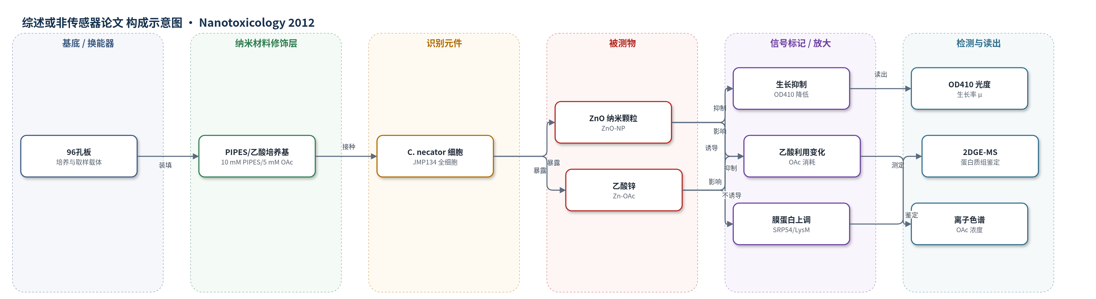
点击图片查看大图 · 依据论文自动绘制
传感器类型
综述或非传感器论文
检测对象
锌氧化物纳米颗粒(ZnO-NPs)、乙酸锌/水合锌离子(Zn-OAc/Zn2+);样品基质:PIPES/乙酸细菌培养基(模拟环境水样/生物介质)
检测原理
本文未构建传统换能器,而是以全细胞作为生物响应元件。Cupriavidus necator 暴露于 ZnO-NP 或 Zn-OAc 后,不同形态锌与细胞界面发生相互作用:Zn-OAc 提供生物可利用 Zn2+,低浓度可促进生长和乙酸利用,高浓度则抑制呼吸与代谢;ZnO-NP 悬浮液中自由 Zn2+ 极低,主要通过与细胞膜接触造成膜损伤。膜损伤触发膜相关蛋白和蛋白转运/合成相关蛋白上调,如 SRP54、LysM 蛋白、EF-Tu 等。信号读出包括 OD410 生长率、乙酸消耗速率以及 2-DGE 胶斑密度,经 MS/MS 鉴定差异蛋白。因此,表型与蛋白质组信号随锌形态和浓度变化,可区分离子毒性与纳米颗粒膜损伤机制。
检测灵敏度
原文未报告 LOD、线性范围、灵敏度斜率或相关系数。
效应效果
在 25 nM–100 mM 总锌范围内,两种锌形态均呈剂量依赖抑制:高于 50 mM 无生长,25 mM 时生长率显著低于基线 0.011 h-1;低于 25 mM 无统计差异,但 Zn-OAc 在 100 nM 出现最优生长率 0.013 h-1,而 ZnO-NP 无促进。乙酸利用基线为 4.2 nM-OAc h-1,50 和 25 mM 时降至约 3 nM-OAc h-1;0.1 和 1 mM Zn-OAc 显著高于基线,ZnO-NP 不显著。蛋白质组显示 Zn-OAc 主要上调代谢与调控蛋白,ZnO-NP 主要上调膜相关蛋白,提示 SRP54 和 LysM 蛋白可作为纳米颗粒膜损伤标志物。作者认为该全细胞响应体系可用于开发生物报告/工程化生物传感器。
传感器的构成
- 响应体系:Cupriavidus necator JMP134 全细胞,作为识别与响应元件
- 培养介质:10 mM PIPES 与 5 mM 乙酸钠,提供乙酸代谢底物
- 暴露样品:ZnO-NP 或 Zn-OAc,提供不同形态锌刺激
- 表型读出:OD410 生长曲线与乙酸消耗,反映细胞活性
- 分子读出:2-DGE 胶斑密度与 MS/MS,鉴定差异表达蛋白
中文摘要
合成金属氧化物纳米材料的毒性通常通过两种机制发挥:一是释放具有毒性的自由金属离子,二是通过表面介导的理化过程产生羟基自由基等活性氧,破坏细胞膜和细胞器。从监管角度看,在生物或自然介质中同时存在纳米颗粒时检测自由金属离子十分困难,使摄取途径和毒性机制难以判定。尽管体外研究表明氧化锌纳米颗粒对土壤细菌Cupriavidus necator的毒性方式与乙酸锌相似,但作者发现纳米颗粒悬浮液中并未检测到自由锌离子。对等浓度纳米颗粒或乙酸锌处理细胞的蛋白质组分析显示,两种锌形态的毒性模式明显不同:纳米颗粒暴露使多种膜相关蛋白上调。数据表明,纳米颗粒主要通过破坏细胞膜导致细胞死亡,而非单纯发挥离子毒性。研究还鉴定出可用于膜损伤毒理基因组学研究的潜在生物标志物基因,或可用于构建工程化生物传感器菌株。
英文摘要
Synthetic metal oxide nanomaterials exert toxicity via two general mechanisms: release of free ions at concentrations which exert toxic effects upon the target cell, or specific surface-mediated physicochemical processes leading to the formation of hydroxyl free radicals and other reactive oxygen species which act to disrupt cell membranes and organelles. From a regulatory standpoint this presents a potential problem since it is not trivial to detect free metal ions in the presence of nanoparticles in biological or natural media. This makes efforts to identify the route of uptake difficult. Although in vitro studies of zinc oxide nanoparticles suggest that toxicity to the soil bacterium Cupriavidus necator is exerted in a similar manner to zinc acetate, we found no free Zn ion is associated with nanoparticle suspensions. The proteome of cells subjected to equal concentrations of either the nanoparticle or zinc acetate suggest that the mode of toxicity is quite different for the two forms of Zn, with a number of membrane-associated proteins up-expressed in response to nanoparticle exposure. Our data suggests that nanoparticles act to interrupt cell membranes thereby causing cell death rather than exerting a strictly toxic effect. We also identify potentially useful genes to serve as biomarkers of membrane disruption in toxicogenomic studies with nanoparticles or to engineer biosensor organisms.