组成图示
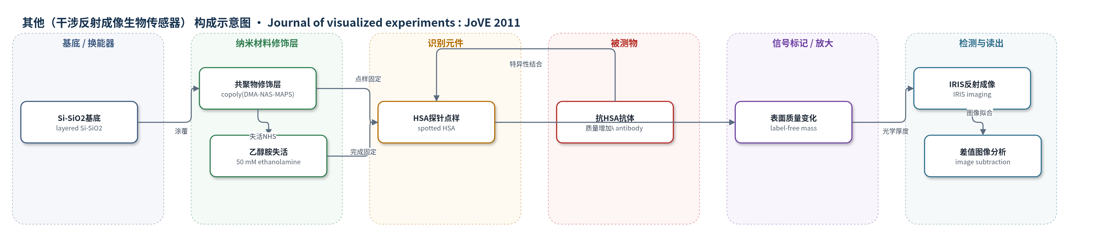
点击图片查看大图 · 依据论文自动绘制
传感器类型
其他(干涉反射成像生物传感器)
检测对象
抗人血清白蛋白抗体(anti-HSA antibody)、Fur 蛋白(Fur protein);样品基质:PBS 缓冲液/溶液
检测原理
IRIS 以层状 Si-SiO2 基底为反射干涉成像表面,copoly(DMA-NAS-MAPS) 修饰层通过表面化学共价固定 HSA、抗体或 DNA 探针。目标分子(如 anti-HSA 抗体或 Fur 蛋白)与识别元件特异性结合后,表面吸附质量增加,使局部光学路径长度/光学厚度改变。IRIS 通过干涉反射成像采集孵育前后的强度图像,经软件拟合得到各点光学厚度,并以点内与背景环的平均差值定量结合质量。信号大小随目标浓度升高而增大;无标记直接检测,不依赖荧光或酶放大,必要时可用二级抗体夹心增加结合质量。
检测灵敏度
原文未报告 LOD、线性范围、灵敏度斜率或相关系数。
效应效果
IRIS 被描述为具有高通量、多路成像、实时与终点测量、试剂消耗少和检测时间短等优点,并采用低成本硅基固相组件,兼容多种表面化学。代表性实验中,500 ng/mL anti-HSA 孵育后,HSA 点样平均光学高度变化为 2.05 nm,而兔 IgG 对照点为 0.13 nm,显示对目标抗体的选择性结合。Fur 蛋白结合随 50、100、200 nM 浓度增加而增强,且较长 dsDNA 探针结合更多;200 nM 时二聚体结合显著增加,提示存在较高非特异结合。原文未报告 RSD、回收率或与 ELISA/HPLC/qPCR 的定量对比。
传感器的构成
- 基底/换能器:layered silicon-SiO2(层状硅-二氧化硅)基底,提供反射干涉成像表面并承载探针阵列
- 表面修饰层:self-adsorbing copoly(DMA-NAS-MAPS)(自吸附共聚物 DMA-NAS-MAPS),提供共价固定探针的表面化学并降低非特异结合
- 识别元件:spotted HSA(人血清白蛋白)或其他抗体/抗原/ssDNA/dsDNA/RNA 探针微阵列,用于特异性捕获目标分子
- 失活/封闭层:50 mM ethanolamine(乙醇胺)用于失活剩余 NHS 基团;可选 BSA/casein/milk 封闭,但本方法未使用
- 信号标记物:label-free(无标记),不添加荧光、酶或比色标记,直接通过结合质量改变光学厚度
- 读出系统:IRIS(interferometric reflectance imaging sensor,干涉反射成像传感器),采集孵育前后图像并计算光学校正厚度/质量变化
中文摘要
生物分子相互作用的灵敏测量在基础生物学、微生物学、环境/农业/生物防御监测、纳米生物技术和诊断等领域具有重要价值。诊断中可通过检测患者样本中目标蛋白或基因组生物标志物的存在、缺失或异常表达来指导治疗;研究中则用于表征分子亲和力与特异性。现有 ELISA、PCR、凝胶电泳和质谱等方法多依赖荧光、放射性或比色标记,存在成本、试剂稳定性、储存安全、标记耗时、批次差异以及标记可能影响蛋白结构和功能等问题,且难以实时直接测量。本文介绍一种适用于微阵列研究的新型无标记光学生物传感器——干涉反射成像传感器(IRIS),可检测蛋白、DNA、抗原材料、完整病毒颗粒及其他生物材料。IRIS 已表现出高灵敏度、高精确性和高重复性,并具备多路成像、实时与终点测量、试剂消耗少、检测时间短等高通量优点。该平台操作简便、设备成本低,采用硅基固相检测组件,兼容多种表面化学方法。本文从探针阵列制备、孵育、目标结合测量到终点结果分析,以 HSA 点样基底捕获抗 HSA 抗体为模型系统展示 IRIS 的使用流程。
英文摘要
The sensitive measurement of biomolecular interactions has use in many fields and industries such as basic biology and microbiology, environmental/agricultural/biodefense monitoring, nanobiotechnology, and more. For diagnostic applications, monitoring (detecting) the presence, absence, or abnormal expression of targeted proteomic or genomic biomarkers found in patient samples can be used to determine treatment approaches or therapy efficacy. In the research arena, information on molecular affinities and specificities are useful for fully characterizing the systems under investigation. Many of the current systems employed to determine molecular concentrations or affinities rely on the use of labels. Examples of these systems include immunoassays such as the enzyme-linked immunosorbent assay (ELISA), polymerase chain reaction (PCR) techniques, gel electrophoresis assays, and mass spectrometry (MS). Generally, these labels are fluorescent, radiological, or colorimetric in nature and are directly or indirectly attached to the molecular target of interest. Though the use of labels is widely accepted and has some benefits, there are drawbacks which are stimulating the development of new label-free methods for measuring these interactions. These drawbacks include practical facets such as increased assay cost, reagent lifespan and usability, storage and safety concerns, wasted time and effort in labelling, and variability among the different reagents due to the labelling processes or labels themselves. On a scientific research basis, the use of these labels can also introduce difficulties such as concerns with effects on protein functionality/structure due to the presence of the attached labels and the inability to directly measure the interactions in real time. Presented here is the use of a new label-free optical biosensor that is amenable to microarray studies, termed the Interferometric Reflectance Imaging Sensor (IRIS), for detecting proteins, DNA, antigenic material, whole pathogens (virions) and other biological material. The IRIS system has been demonstrated to have high sensitivity, precision, and reproducibility for different biomolecular interactions [1-3]. Benefits include multiplex imaging capacity, real time and endpoint measurement capabilities, and other high-throughput attributes such as reduced reagent consumption and a reduction in assay times. Additionally, the IRIS platform is simple to use, requires inexpensive equipment, and utilizes silicon-based solid phase assay components making it compatible with many contemporary surface chemistry approaches. Here, we present the use of the IRIS system from preparation of probe arrays to incubation and measurement of target binding to analysis of the results in an endpoint format. The model system will be the capture of target antibodies which are specific for human serum albumin (HSA) on HSA-spotted substrates.