组成图示
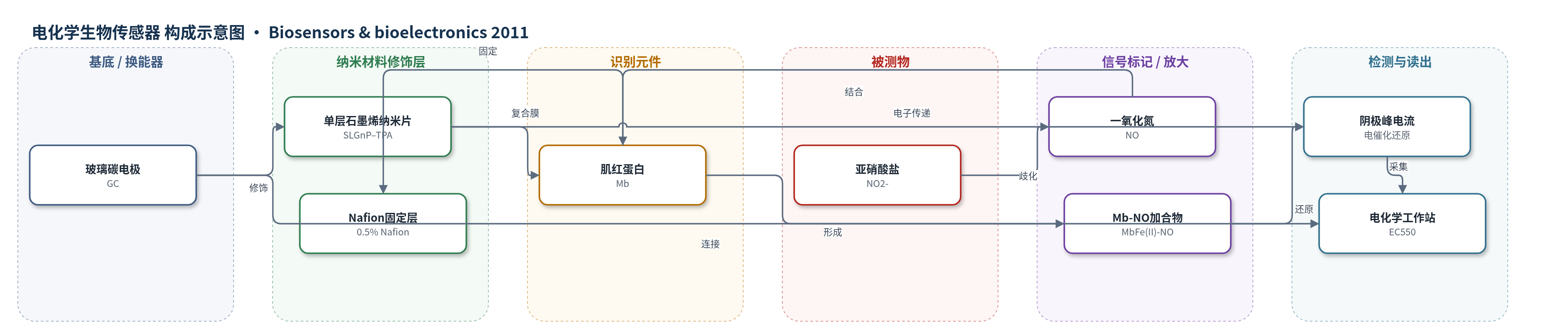
点击图片查看大图 · 依据论文自动绘制
传感器类型
电化学生物传感器
检测对象
亚硝酸盐(nitrite, NO2-);样品基质:pH 5.0 磷酸盐缓冲液(文中亦提及食品、饮用水、环境样品)
检测原理
在酸性介质中,亚硝酸盐发生歧化反应生成一氧化氮:3NO2- + H2O ⇌ 2NO + NO3- + 2OH-。NO 与 Mb 血红素 Fe(II) 结合形成 MbFe(II)–NO。SLGnP 作为电子导线缩短 Mb 血红素中心与 GC 电极间的电子转移距离,使 MbFe(III)/Fe(II) 发生直接电子转移。随后 MbFe(II)–NO 在电极表面被电催化还原:MbFe(II)–NO + 2e- + 2H+ → MbFe(II) + H2O + N2O。阴极峰电流随 NO2- 浓度增加而增大,从而实现亚硝酸盐的间接电化学检测。
检测灵敏度
LOD: 0.01 mM;线性范围: 0.05–2.5 mM;灵敏度: 3.42 × 10^4 μA M−1 cm−2;线性方程: I/μA = 0.198 + 0.598 C/mM;R = 0.996 (n=9)
效应效果
该传感器在 pH 5.0 磷酸盐缓冲液中表现出良好稳定性:200 次连续循环伏安扫描后电流保持恒定;4 ℃ 保存约 2 周仍保留约 90% 初始灵敏度。对 0.1 mM NO2- 的 8 次连续测量 RSD 为 4.1%,6 个同批制备电极的批间 RSD 为 5.2%。抗干扰方面,尿酸浓度超过 NO2- 10 倍时未观察到电流响应变化。与 Hb–丝素蛋白膜(LOD 0.233 mM)和 Hb/CTAB(LOD 0.286 mM)相比,本传感器 LOD 0.01 mM 具有竞争力。作者认为其可用于食品、饮用水和环境样品中亚硝酸盐的快速、原位监测。
传感器的构成
- 基底/换能器电极:玻璃碳电极(GC),经氧化铝抛光和清洗,作为电子转移动态基底。
- 纳米材料修饰层:单层石墨烯纳米片–四钠1,3,6,8-芘四磺酸(SLGnP–TPA),提供导电网络、生物相容微环境和电子导线。
- 识别/催化元件:肌红蛋白(Mb),与 SLGnP–TPA 静电作用形成复合膜,保留天然结构并催化 NO 还原。
- 固定/结合层:Nafion(0.5%),滴涂于复合膜表面作为 binder,防止膜脱落并稳定电极。
- 信号标记物:无外源标记,Mb 血红素 Fe(III)/Fe(II) 直接电化学及电催化 NO 还原产生阴极电流。
中文摘要
本文报道了一种基于单层石墨烯纳米片(SLGnP)–蛋白复合膜的新型亚硝酸盐生物传感器。SLGnP 具有优异的生物相容性、导电性和对局部扰动的敏感性,可为蛋白固定提供生物相容微环境,并在血红素蛋白电活性中心与电极表面之间提供合适的电子转移距离。在复合膜修饰电极上,获得一对定义良好且准可逆的循环伏安峰,反映肌红蛋白(Mb)三价/二价铁偶联的直接电化学。场发射扫描电镜(FESEM)和紫外–可见光谱(UV–vis)表征表明,复合膜具有独特形貌,且其中蛋白保留了类似天然态的二级结构。该复合膜对一氧化氮(NO)还原表现出优异的电催化能力,并用于间接测定亚硝酸盐。传感器对亚硝酸盐具有良好的电化学响应,线性范围为 0.05–2.5 mM,检出限为 0.01 mM。
英文摘要
A novel nitrite biosensor was developed through a sensing platform consisted of single-layer graphene nanoplatelet (SLGnP)-protein composite film. SLGnP with the virtues of excellent biocompatibility, conductivity and high sensitivity to the local perturbations can provide a biocompatible microenvironment for protein immobilization and a suitable electron transfer distance between electroactive centers of heme protein and electrode surface. A pair of well-defined and quasi-reversible cyclic voltammetric peaks that reflected the direct electrochemistry for ferric/ferrous couple of myoglobin (Mb) was achieved at the composite film modified electrode. Field emission scanning electron microscopy (FESEM) and ultraviolet visible spectra (UV-vis) were utilized to characterize the composite film. The results demonstrated that the morphology of the composite film was unique and the protein in the composite film retained its secondary structure similar to the native state. The composite film also displayed excellent electrocatalytic ability for the reduction of nitric oxide, which was applied to determine nitrite indirectly. It exhibited good electrochemical response to nitrite with a linear range from 0.05 to 2.5 mM and a detection limit of 0.01 mM.