组成图示
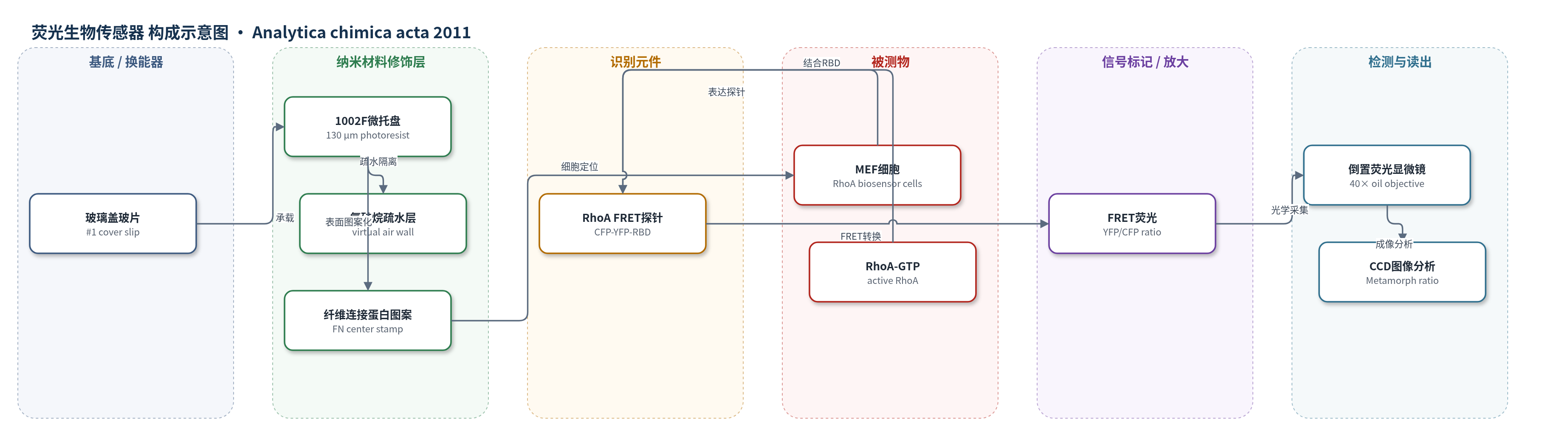
点击图片查看大图 · 依据论文自动绘制
传感器类型
荧光生物传感器
检测对象
细胞内RhoA GTPase活性(RhoA-GTP);样品基质为贴壁培养的小鼠胚胎成纤维细胞(MEF)及细胞培养液(Ham's F-12K,2% FBS,10 mM HEPES)。
检测原理
该检测基于活细胞内RhoA FRET生物传感器。MEF细胞表达CFP-YFP-RBD融合探针,CFP为供体,YFP为受体,RBD为RhoA结合域。当RhoA装载GTP成为活性RhoA-GTP时,RBD与RhoA结合,使CFP与YFP的相对取向和距离改变,FRET效率升高;在CFP激发下YFP发射增强,YFP/CFP荧光比随局部RhoA活性增加而升高。纤维连接蛋白中心图案将细胞主体定位在托盘中心,远离粗糙侧壁,降低边缘散射光干扰。高NA倒置荧光显微镜分别采集CFP与YFP图像,经配准、漂白校正和比值计算,获得空间分辨的RhoA活性分布。该体系未使用酶促或核酸放大,信号直接来自FRET效率变化。
检测灵敏度
未报告LOD、线性范围、灵敏度斜率或相关系数。
效应效果
中心纤维连接蛋白图案显著提高细胞定位精度:H1299细胞中心定位率为80±7%,全表面涂覆对照为21±6%;MEF细胞中心定位率为85±4%,对照为25±3%;HeLa细胞结果类似。10,000个托盘阵列上30 μm圆图案中心偏差为13±8%(n=10),连续印刷偏差为11±9%。中心图案使细胞荧光与托盘边缘散射光分离,在0.33 μm空间分辨率和30 s时间间隔下观察到细胞伪足边缘RhoA激活及伸出/回缩周期。作者认为该平台可扩展细胞筛选标准,用于基于动态信号特性的细胞分离。
传感器的构成
- 基底/支撑层:玻璃盖玻片(#1 cover slip,约150 μm),承载微托盘并适配高NA油镜工作距离。
- 微托盘承载层:1002F光刻胶微托盘(130 μm×130 μm,高25或50 μm),提供可释放单元并隔离单个细胞。
- 疏水隔离层:十七氟-1,1,2,2-四氢癸基三氯硅烷气相沉积,形成虚拟空气墙,减少托盘间细胞附着。
- 细胞定位层:纤维连接蛋白(fibronectin, FN)中心微图案(90 μm×90 μm),引导细胞贴附于托盘中心。
- 传感细胞层:表达RhoA FRET生物传感器的MEF细胞,作为活细胞传感平台。
- 识别/信号元件:CFP-YFP-RBD融合RhoA探针,RhoA-GTP结合RBD改变CFP与YFP取向。
- 光学读出层:倒置荧光显微镜、40×油镜、CCD相机与滤光片,采集CFP/YFP荧光并计算FRET比。
中文摘要
托盘阵列可在细胞保持贴壁状态下实现分离,并拓宽细胞筛选标准,但仍需纳入动态细胞内信号事件。为证明在阵列上用高分辨显微成像测量细胞蛋白行为的可行性,作者修饰单个托盘表面以减少边缘散射光,并用定制印章工具将纤维连接蛋白以不同形状和尺寸的微图案印在三维托盘表面。中心纤维连接蛋白图案使H1299和小鼠胚胎成纤维细胞(MEF)定位到托盘中心并远离边缘,与全表面涂覆相比,中心定位细胞比例提高3–4倍,同时使细胞荧光与粗糙侧壁散射光分离。作者进一步用RhoA GTPase生物传感器进行高空间分辨率荧光测量,该传感器通过青色荧光蛋白(CFP)与黄色荧光蛋白(YFP)的荧光共振能量转移(FRET)检测细胞边缘伪足中的局部RhoA活性。结果表明,可在托盘阵列上实现荧光传感器的空间分辨测量,有望基于复杂细胞信号特性进行新型细胞分离。
英文摘要
Pallet arrays enable cells to be separated while they remain adherent to a surface and provide a much greater range of cell selection criteria relative to that of current technologies. However there remains a need to further broaden cell selection criteria to include dynamic intracellular signaling events. To demonstrate the feasibility of measuring cellular protein behavior on the arrays using high resolution microscopy, the surfaces of individual pallets were modified to minimize the impact of scattered light at the pallet edges. The surfaces of the three-dimensional pallets on an array were patterned with a coating such as fibronectin using a customized stamping tool. Micropatterns of varying shape and size were printed in designated regions on the pallets in single or multiple steps to demonstrate the reliability and precision of patterning molecules on the pallet surface. Use of a fibronectin matrix stamped at the center of each pallet permitted the localization of H1299 and mouse embryonic fibroblast (MEF) cells to the pallet centers and away from the edges. Compared to pallet arrays with fibronectin coating the entire top surface, arrays with a central fibronectin pattern increased the percentage of cells localized to the pallet center by 3-4-fold. Localization of cells to the pallet center also enabled the physical separation of cells from optical artifacts created by the rough pallet side walls. To demonstrate the measurement of dynamic intracellular signaling on the arrays, fluorescence measurements of high spatial resolution were performed using a RhoA GTPase biosensor. This biosensor utilized fluorescence resonance energy transfer (FRET) between cyan fluorescent protein (CFP) and yellow fluorescent protein (YFP) to measure localized RhoA activity in cellular ruffles at the cell periphery. These results demonstrated the ability to perform spatially resolved measurements of fluorescence-based sensors on the pallet arrays. Thus, the patterned pallet arrays should enable novel cell separations in which cell selection is based on complex cellular signaling properties.