组成图示
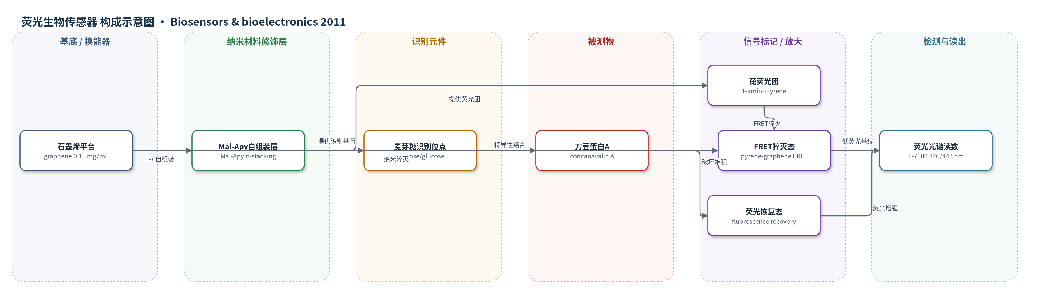
点击图片查看大图 · 依据论文自动绘制
传感器类型
荧光生物传感器
检测对象
刀豆蛋白A(ConA, concanavalin A);样品基质:PBS缓冲液(10 mM, pH 7.0,含0.1 mM MnCl2和0.1 mM CaCl2)
检测原理
Mal-Apy由1-氨基芘与麦芽糖经还原胺化连接,芘环提供荧光,麦芽糖/葡萄糖基团识别ConA。Mal-Apy通过芘环与石墨烯的π-π堆积自组装到石墨烯表面,芘环与石墨烯距离接近,发生FRET,石墨烯作为纳米淬灭剂使荧光猝灭。加入ConA后,ConA与麦芽糖/葡萄糖基团特异性结合,改变Mal-Apy构象或将其从石墨烯表面解离,破坏π-π堆积,使芘环远离石墨烯,FRET效率下降,荧光恢复。ConA浓度越高,结合越多,荧光恢复越强,在447 nm发射处监测相对荧光强度即可定量。检测无需洗涤或分离,5 min达到平衡。
检测灵敏度
LOD: 0.8 nM;线性范围: 2.0 × 10⁻² to 1.0 μM;线性方程: y = 1.029x + 0.284;斜率: 1.029;R = 0.996
效应效果
该石墨烯FRET平台对ConA具有选择性,BSA在0–5.0 μM范围内仅引起较低荧光响应,而ConA可显著增强荧光,说明麦芽糖/葡萄糖基团对ConA识别特异。石墨烯猝灭效率重现性良好,三次平行实验中Mal-Apy剩余荧光分别为14.7%、12.1%和12.2%,标准偏差约1.2%;ConA检测的相对标准偏差为7.9%。检测在5 min内达到平衡,LOD 0.8 nM,与已报道最佳结果相当。方法为同相检测,无需洗涤、离心或分离,适合快速、灵敏、选择性检测ConA,并可拓展至其他凝集素、药物筛选、双分子识别和疾病诊断。
传感器的构成
- 纳米材料/信号平台:石墨烯(graphene),作为π-π堆积载体和FRET纳米淬灭剂,猝灭芘荧光。
- 识别-荧光双功能分子:麦芽糖接枝氨基芘(Mal-Apy),通过芘环自组装到石墨烯表面,同时提供识别基团和荧光团。
- 识别元件:Mal-Apy中的麦芽糖/葡萄糖基团(maltose/glucose),特异性结合ConA。
- 信号标记物:Mal-Apy中的1-氨基芘(1-aminopyrene, 1-AP)芘环,作为荧光团,经FRET猝灭/恢复。
- 反应介质:10 mM PBS缓冲液(pH 7.0,含0.1 mM MnCl2和0.1 mM CaCl2),维持ConA与糖基团结合活性。
中文摘要
本文提出一种基于荧光共振能量转移(FRET)的同相检测刀豆蛋白A(ConA)的新型生物传感器。作者合成并表征了麦芽糖接枝氨基芘(Mal-Apy),将其用于荧光开关和ConA识别。Mal-Apy通过π-π堆积作用自组装到石墨烯表面后,由于石墨烯作为芘环的“纳米淬灭剂”发生FRET,其荧光被显著猝灭。当存在ConA时,ConA与葡萄糖基团的竞争性结合破坏芘环与石墨烯之间的π-π堆积作用,使荧光恢复。该方法表现出对ConA的选择性检测,线性范围为2.0×10^-2至1.0 μM,线性方程为y=1.029x+0.284(R=0.996),检出限低至0.8 nM,检测可在5 min内完成,说明该方法可用于快速、灵敏、选择性检测ConA。数据表明石墨烯FRET平台在蛋白-碳水化合物相互作用研究中具有潜力,并可广泛应用于药物筛选、双分子识别和疾病诊断。
英文摘要
In this work, we proposed a novel biosensor to homogeneously detect concanavalin A (ConA) using pyrene-conjugated maltose assembled graphene based on fluorescence resonance energy transfer (FRET). Maltose-grafted-aminopyrene (Mal-Apy) was synthesized and characterized by mass spectra, UV-vis and fluorescence spectra. The Mal-Apy was further employed for fluorescence switch and ConA recognition. When Mal-Apy was self-assembled on the surface of graphene by means of π-stacking interaction, its fluorescence was adequately quenched because the graphene acted as a "nanoquencher" of the pyrene rings due to FRET. As a result, in the presence of ConA, competitive binding of ConA with glucose destroyed the π-stacking interaction between the pyrene and graphene, thereby causing the fluorescence recovery. This method was demonstrated the selective sensing of ConA, and the linear range is 2.0 × 10⁻² to 1.0 μM with the linear equation y=1.029x + 0.284 (R = 0.996). The limit of detection for ConA was low to 0.8 nM, and the detection of ConA could be performed in 5 min, indicating that this method could be used for fast, sensitive, and selective sensing of ConA. Such data suggests that the graphene FRET platform is a great potential application for protein-carbohydrate studies, and would be widely applied in drug screening, bimolecular recognition and disease diagnosis.