组成图示
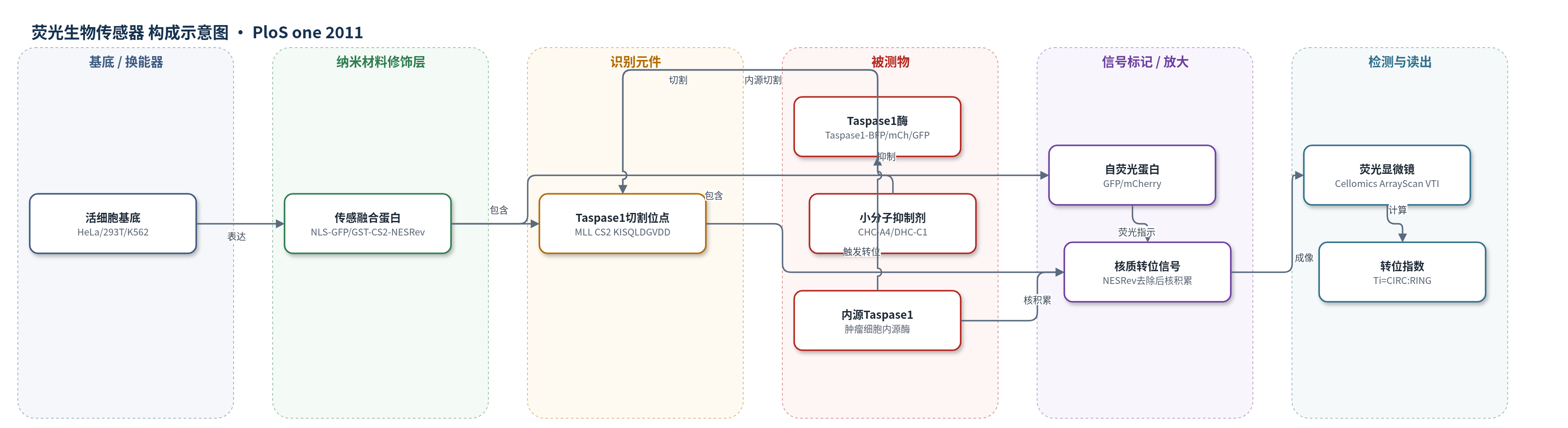
点击图片查看大图 · 依据论文自动绘制
传感器类型
荧光生物传感器
检测对象
Taspase1 蛋白酶活性(Taspase1 protease activity)、Taspase1 小分子抑制剂(Taspase1 inhibitors);样品基质:活细胞(HeLa、293T、K562 及肿瘤细胞系)
检测原理
传感融合蛋白含核输入信号(NLS)、GST、Taspase1切割位点、自荧光蛋白和核输出信号(NESRev)。未切割时NESRev主导,使GFP/mCherry主要位于胞质。当Taspase1识别P1位天冬氨酸并切割Myc-NESRev后,核输出信号被移除,NLS主导使荧光蛋白进入细胞核,核/胞质荧光比升高。Taspase1活性越高或抑制剂越少,核积累越强;小分子抑制剂结合Taspase1活性中心后降低切割效率,转位指数下降。该体系以蛋白酶切割为识别事件,以核质分布变化为换能机制,通过荧光显微镜定量读出,无需外源标记或酶促放大。
检测灵敏度
原文未报告 LOD、线性范围、灵敏度斜率或 R^2;仅报告高通量筛选 Z’factor = 0.63,初筛判据 Tic > 2。
效应效果
该传感器对Taspase1具有良好选择性:失活突变TaspT234V、Caspase3/9及Rev不引起核积累,LMB处理可排除核输入干扰。活细胞检测在酶/底物1:10时仍高效,优于体外重组体系。高通量平台Z’factor=0.63,筛选聚焦化合物库和90种真菌提取物,发现CHC-A4与DHC-C1在50 μM时部分抑制转位,5 μM无抑制,微注射增强抑制,初筛命中率约3%。体系可检测实体瘤细胞内源Taspase1,并验证FRM4B、PTRZ、DPOLZ等新底物,作者认为其适用于高内涵筛选Taspase1抑制剂。
传感器的构成
- 细胞基底:活细胞(HeLa、293T、K562 及多种肿瘤细胞系)作为表达、核质转运与蛋白酶反应环境
- 传感融合蛋白:NLS-GFP/GST-CS2-NESRev(TS-Cl2+)或 NLS-mCh/GST-CS2-NESRev(TS-Cl2+R),将切割事件转化为核质荧光分布变化
- 识别元件:Taspase1 切割位点 CS2(KISQLDGVDD)或 FRM4B/PTRZ/DPOLZ/TFIIA 切割位点,被 Taspase1 特异性识别并切割
- 信号标记物:GFP 或 mCherry 自荧光蛋白,切割后核内积累产生荧光信号
- 定位调控元件:SV40 大 T 抗原核输入信号(NLS)与 HIV-1 Rev 核输出信号(NESRev),未切割时使传感蛋白主要定位于胞质
- 酶/靶标:Taspase1(Taspase1-BFP、Taspase1-mCh、Taspase1-GFP 或内源 Taspase1),催化传感蛋白切割
- 读出平台:Cellomics ArrayScan VTI 荧光显微镜与 Hoechst 33342 核染色,计算核/胞质荧光比(Ti)
中文摘要
背景:苏氨酸天冬氨酸蛋白酶1(Taspase1)可切割混合性白血病(MLL)蛋白及致白血病MLL融合蛋白,但缺乏小分子抑制剂和细胞功能检测体系。方法/结果:本研究建立了用于在活细胞内探测Taspase1功能的高效细胞生物传感器。该传感器由谷胱甘肽S-转移酶(GST)、自荧光蛋白变体、Taspase1切割位点以及核输入/输出信号组合构成。未切割时传感器主要定位于胞质;表达有活性的Taspase1而非失活突变体或Caspase3时,可触发其切割并使荧光蛋白核内积累。与重组蛋白体外检测相比,活细胞检测效率更高。基于优化的核转位算法,三色检测可适配高通量显微平台(Z’factor=0.63)。自动高内涵分析用于筛选经虚拟药效团筛选获得的聚焦化合物库及真菌提取物,发现两种化合物可部分抑制活细胞中Taspase1切割。该体系还可用于检测实体瘤细胞模型中的内源Taspase1,并鉴定更高效的Taspase1切割共识序列,进而预测并验证FRM4B、PTRZ和DPOLZ等新底物。结论:该检测体系可用于遗传学探测Taspase1结构功能,并适用于高内涵筛选Taspase1抑制剂。
英文摘要
BACKGROUND: Threonine Aspartase 1 (Taspase1) mediates cleavage of the mixed lineage leukemia (MLL) protein and leukemia provoking MLL-fusions. In contrast to other proteases, the understanding of Taspase1's (patho)biological relevance and function is limited, since neither small molecule inhibitors nor cell based functional assays for Taspase1 are currently available.
METHODOLOGY/FINDINGS: Efficient cell-based assays to probe Taspase1 function in vivo are presented here. These are composed of glutathione S-transferase, autofluorescent protein variants, Taspase1 cleavage sites and rational combinations of nuclear import and export signals. The biosensors localize predominantly to the cytoplasm, whereas expression of biologically active Taspase1 but not of inactive Taspase1 mutants or of the protease Caspase3 triggers their proteolytic cleavage and nuclear accumulation. Compared to in vitro assays using recombinant components the in vivo assay was highly efficient. Employing an optimized nuclear translocation algorithm, the triple-color assay could be adapted to a high-throughput microscopy platform (Z'factor = 0.63). Automated high-content data analysis was used to screen a focused compound library, selected by an in silico pharmacophor screening approach, as well as a collection of fungal extracts. Screening identified two compounds, N-[2-[(4-amino-6-oxo-3H-pyrimidin-2-yl)sulfanyl]ethyl]benzenesulfonamide and 2-benzyltriazole-4,5-dicarboxylic acid, which partially inhibited Taspase1 cleavage in living cells. Additionally, the assay was exploited to probe endogenous Taspase1 in solid tumor cell models and to identify an improved consensus sequence for efficient Taspase1 cleavage. This allowed the in silico identification of novel putative Taspase1 targets. Those include the FERM Domain-Containing Protein 4B, the Tyrosine-Protein Phosphatase Zeta, and DNA Polymerase Zeta. Cleavage site recognition and proteolytic processing of these substrates were verified in the context of the biosensor.
CONCLUSIONS: The assay not only allows to genetically probe Taspase1 structure function in vivo, but is also applicable for high-content screening to identify Taspase1 inhibitors. Such tools will provide novel insights into Taspase1's function and its potential therapeutic relevance.