组成图示
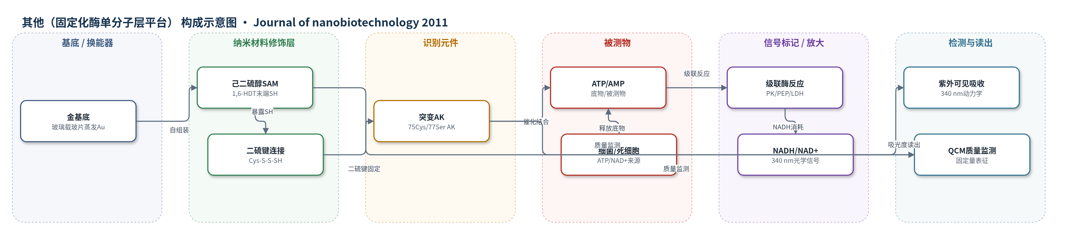
点击图片查看大图 · 依据论文自动绘制
传感器类型
其他(固定化酶单分子层平台)
检测对象
腺苷三磷酸(ATP)、腺苷一磷酸(AMP)、烟酰胺腺嘌呤二核苷酸(NAD+);样品基质:反应缓冲液(TRIS/imidazole-acetate buffer),概念样品为细菌或死细胞释放物。
检测原理
突变AK通过75位Cys与HDT SAM末端SH自发形成S-S键,使蛋白以活性域朝外的取向固定,77位Ser避免额外Cys干扰。加入ATP/AMP后,AK催化磷酸基团从Mg-ATP转移到AMP生成ADP;在级联体系中,PK利用PEP再生ATP,LDH消耗NADH生成NAD+,使340 nm处NADH吸光度随时间下降。ATP/AMP或NAD+水平改变级联反应速率,从而改变吸光度斜率。该策略利用定向固定提高活性位点可及性,并通过ADP-ATP循环级联放大信号;QCM仅用于监测固定质量,不作为主要传感读出。
检测灵敏度
未报告LOD、线性范围、相关系数;动力学斜率: -0.0732 至 -0.1198(任意单位),平均 -0.097;溶液对照(52 nmol 蛋白)斜率: -0.1370。
效应效果
固定化AK未损失酶功能,且活性约为相同量溶液AK的100倍;考虑样品尺寸误差后仍至少高90倍。缺失任一反应组分时无催化反应,说明固定化未降低特异性。动力学斜率在-0.0732至-0.1198(任意单位)之间,平均-0.097;复测样本斜率-0.1198与-0.1130在实验误差内,至少5次独立实验。QCM显示蛋白覆盖量与单分子表面积模型一致,覆盖≤60%,留下自由体积利于LID域运动。直接吸附裸金无活性,而HDT SAM二硫键固定可保持高活性。作者认为该平台可用于快速检测细菌感染或死细胞释放的ATP/NAD+,比临床培养2-24小时更快,并拟转移至纳米管高长径比平台。
传感器的构成
- 基底:金(Au)蒸发于玻璃载玻片,厚度约2000 Å,提供支撑与界面。
- 自组装单分子层:1,6-己二硫醇(1,6-hexanedithiol, HDT)在Au上形成SAM,末端暴露SH基团。
- 连接/还原层:TCEP-HCl(tris(2-carboxyethyl)phosphine hydrochloride)还原氧化SH并防止蛋白聚集。
- 识别/催化元件:突变腺苷酸激酶(Adenylate kinase, AK;75Cys/77Ser)经Cys与SAM末端SH形成S-S键定向固定。
- 反应介质:TRIS缓冲液(pH 7.2)用于固定;检测介质含NADH、AMP、PEP、MgCl2、醋酸钾、咪唑/醋酸缓冲液(pH 7.5)。
- 级联酶元件:丙酮酸激酶(PK)和L-乳酸脱氢酶(LDH)与磷酸烯醇式丙酮酸(PEP)构成ADP-ATP/NADH级联反应。
- 信号标记:NADH/NAD+作为光学信号分子,340 nm吸光度变化反映反应动力学。
- 质量换能器:石英晶体微天平(QCM)金覆盖晶体(0.392 cm²),用于监测SAM与AK质量/覆盖量。
中文摘要
本文报道一种用于精确固定化生物分子并保留其天然功能的新方法,以腺苷酸激酶(AK)为模型系统。研究在金表面构建1,6-己二硫醇(HDT)自组装单分子层(SAM),并通过接触角、Elman试剂、石英晶体微天平(QCM)和X射线光电子能谱(XPS)表征。作者设计突变AK,将75位残基替换为半胱氨酸、77位半胱氨酸替换为丝氨酸,使蛋白通过自发形成的二硫键定向连接至SAM末端巯基。XPS显示HDT SAM中存在连接金的硫醇盐硫和末端SH/S-S硫;QCM与XPS共同证实蛋白单分子层形成,且表面蛋白量与单分子表面积模型一致。酶活测试表明固定化AK未损失催化功能,其活性约为相同量溶液AK的100倍。作者认为该平台可用于活性蛋白的图案化定位和bioMEMS,并计划将其转移至高长径比纳米管平台以开发生物传感器。
英文摘要
BACKGROUND: The interest in introducing ecologically-clean, and efficient enzymes into modern industry has been growing steadily. However, difficulties associated with controlling their orientation, and maintaining their selectivity and reactivity is still a significant obstacle. We have developed precise immobilization of biomolecules, while retaining their native functionality, and report a new, fast, easy, and reliable procedure of protein immobilization, with the use of Adenylate kinase as a model system.
METHODS: Self-assembled monolayers of hexane-1,6-dithiol were formed on gold surfaces. The monolayers were characterized by contact-angle measurements, Elman-reagent reaction, QCM, and XPS. A specifically designed, mutated Adenylate kinase, where cysteine was inserted at the 75 residue, and the cysteine at residue 77 was replaced by serine, was used for attachment to the SAM surface via spontaneously formed disulfide (S-S) bonds. QCM, and XPS were used for characterization of the immobilized protein layer. Curve fitting in XPS measurements used a Gaussian-Lorentzian function.
RESULTS AND DISCUSSION: Water contact angle (65-70°), as well as all characterization techniques used, confirmed the formation of self-assembled monolayer with surface SH groups. X-ray photoelectron spectroscopy showed clearly the two types of sulfur atom, one attached to the gold (triolate) and the other (SH/S-S) at the ω-position for the hexane-1,6-dithiol SAMs. The formation of a protein monolayer was confirmed using XPS, and QCM, where the QCM-determined amount of protein on the surface was in agreement with a model that considered the surface area of a single protein molecule. Enzymatic activity tests of the immobilized protein confirmed that there is no change in enzymatic functionality, and reveal activity ~100 times that expected for the same amount of protein in solution.
CONCLUSIONS: To the best of our knowledge, immobilization of a protein by the method presented here, with the resulting high enzymatic activity, has never been reported. There are many potential applications for selective localization of active proteins at patterned surfaces, for example, bioMEMS (MEMS--Micro-Electro-Mechanical Systems. Due to the success of the method, presented here, it was decided to continue a research project of a biosensor by transferring it to a high aspect ratio platform--nanotubes.