组成图示
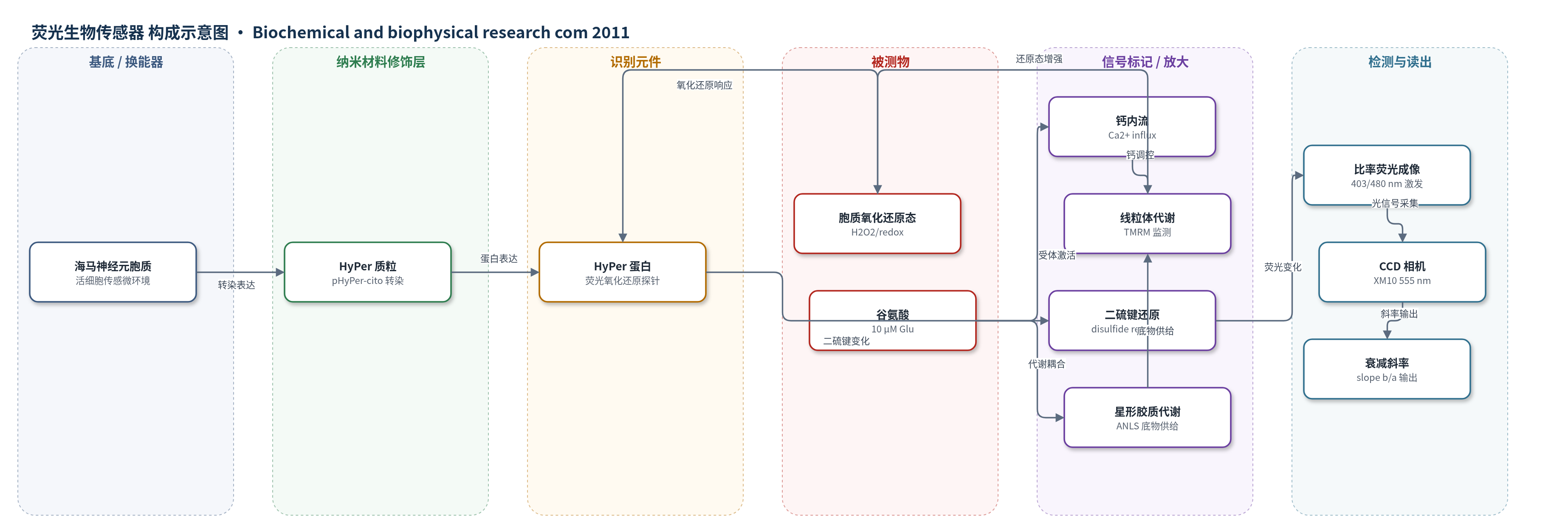
点击图片查看大图 · 依据论文自动绘制
传感器类型
荧光生物传感器
检测对象
胞质氧化还原状态(cytoplasmic redox state,以 HyPer 荧光衰减率表征);样品基质:原代培养大鼠海马神经元胞质(hippocampal neuron cytoplasm),含星形胶质细胞共培养
检测原理
HyPer 是基因编码的荧光氧化还原探针,其半胱氨酸对在氧化环境中形成二硫键,改变比率荧光。实验先用 100 μM H2O2 短脉冲将 HyPer 氧化至平台,随后胞质抗氧化系统将其还原,荧光比率按基础速率衰减。谷氨酸(10 μM)激活 NMDA 受体,引起 Ca2+ 内流,并通过线粒体脱氢酶增强氧化磷酸化;星形胶质细胞经 ANLS 提供乳酸/丙酮酸,增加胞质还原当量,使 HyPer 二硫键还原加速,衰减斜率增大。信号由 403/12 nm 与 480/20 nm 激发、555/28 nm 发射的比率荧光成像读出,以斜率 b/a 反映胞质还原态增强。
检测灵敏度
基础衰减率: 0.04 ± 0.01 min^-1;谷氨酸使 HyPer 衰减斜率平均增加约 7 倍
效应效果
谷氨酸效应约在 80% 神经元中出现,平均使 HyPer 衰减斜率增加约 7 倍;NMDA(200 μM)和 D-Asp(10 μM)可模拟该效应,AP-5 与 DNQX 可阻断。无钙加 EGTA 消除效应,恢复 Ca2+ 后重现;veratridine(75 μM)、monensin(100 μM)、ionomycin(2 μM)均可加速衰减,提示 Ca2+ 内流关键。抗霉素 A(0.25 μg/ml)和氟乙酸盐(5 mM)分别阻断线粒体与星形胶质代谢后效应消失;乳酸/丙酮酸可加速衰减。pH 下降 0.1 单位不足以解释信号,0.5 单位才引起 0.8 比率下降。作者认为该氧化还原偏移可保护神经元兴奋传递,并为谷氨酸能代谢耦合提供实时成像方法。
传感器的构成
- 传感载体:pHyPer-cito 质粒,经 Lipofectamine 2000 或磷酸钙转染,在神经元胞质表达 HyPer
- 识别元件:HyPer 蛋白中的半胱氨酸对(Cys pairs),响应胞质 H2O2/氧化还原状态并形成或还原二硫键
- 信号标记物:HyPer 比率荧光,403/12 nm 与 480/20 nm 激发、555/28 nm 发射,氧化/还原态改变衰减率
- 样品基质:原代培养大鼠海马神经元(hippocampal neurons)与星形胶质细胞(astrocytes)共培养,KRH 缓冲液
- 光学换能/读出:Olympus IX81 倒置显微镜、40×物镜、MT20E 发射轮、XM10 CCD 相机,实时成像
- 氧化预处理:H2O2 100 μM 短脉冲,将 HyPer 氧化至平台,便于监测后续还原速率
中文摘要
脑细胞代谢与细胞内氧化还原(redox)平衡密切相关。谷氨酸能传递伴随神经元底物偏好改变。为此,作者研究培养海马神经元暴露于谷氨酸时的胞质氧化还原变化。神经元转染 HyPer,一种用于过氧化氢的基因编码氧化还原生物传感器,可实时成像氧化还原状态。与生物传感器还原相对应的荧光衰减速率,在低剂量谷氨酸(10 μM)以及药理学刺激 NMDA 谷氨酸受体时均增强。急性螯合细胞外 Ca2+ 可消除谷氨酸对 HyPer 荧光的影响。进一步实验表明,线粒体功能及能量底物可用性决定神经元氧化还原状态,并且是谷氨酸影响生物传感器信号所必需的。此外,结果提示星形胶质细胞代谢参与谷氨酸所诱导的神经元氧化还原状态变化。
英文摘要
Brain cell metabolism is intimately associated with intracellular oxidation-reduction (redox) balance. Glutamatergic transmission is accompanied with changes in substrate preference in neurons. Therefore, we studied cytoplasmatic redox changes in hippocampal neurons in culture exposed to glutamate. Neurons were transfected with HyPer, a genetically encoded redox biosensor for hydrogen peroxide which allows real-time imaging of the redox state. The rate of fluorescence decay, corresponding to the reduction of the biosensor was found to be augmented by low doses of glutamate (10 μM) as well as by pharmacological stimulation of NMDA glutamate receptors. Acute chelation of extracellular Ca(2+) abolished the glutamate-induced effect observed on HyPer fluorescence. Additional experiments indicated that mitochondrial function and hence energetic substrate availability commands the redox state of neurons and is required for the glutamate effect observed on the biosensor signal. Furthermore, our results implicated astrocytic metabolism in the changes of neuronal redox state observed with glutamate.