组成图示
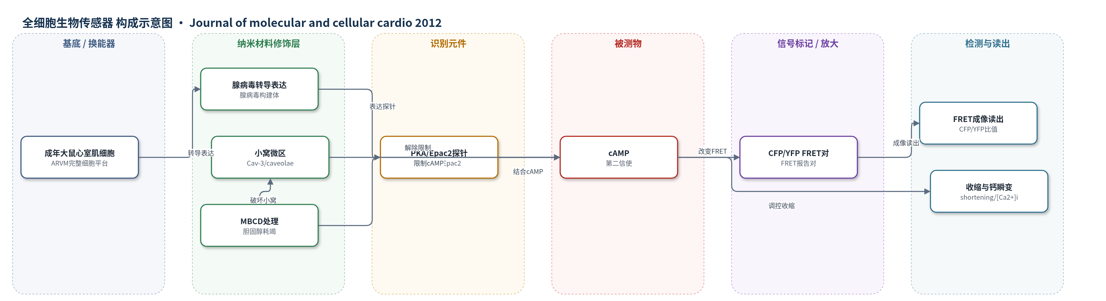
点击图片查看大图 · 依据论文自动绘制
传感器类型
全细胞生物传感器
检测对象
环磷酸腺苷(cAMP),样品基质为完整成年大鼠心室肌细胞(ARVM)内PKA-RII区室与细胞质。
检测原理
该检测基于FRET cAMP生物传感器。腺病毒将PKA-RII或Epac2基cAMP探针转导至成年大鼠心室肌细胞(ARVM);探针含CFP/YFP荧光对,cAMP结合PKA-RII或Epac2 cAMP结合域后引起构象变化,改变CFP供体向YFP受体的荧光共振能量转移效率,以CFP/YFP比值实时反映局部cAMP浓度。PKA探针经AKAP锚定于PKA-RII区室,Epac2探针报告细胞质cAMP。β2-肾上腺素受体(β2-AR)激动后通过Gs/Gi调控腺苷酸环化酶(AC5/6)产生cAMP;caveolae/Cav-3微区限制cAMP产生与传播。MBCD耗竭胆固醇破坏caveolae,使PKA-RII区室cAMP升高,FRET响应增强,从而显示cAMP从小窝约束中释放。
检测灵敏度
原文未报告LOD、线性范围、灵敏度斜率或相关系数。
效应效果
PKA探针在控制细胞ZNT刺激仅1.3±1.4% FRET变化,MBCD后4.7±1.4%,显示区室选择性;Epac2探针控制与MBCD分别为4.3±0.8%和4.2±0.5%,说明细胞质cAMP未变。MBCD使ZNT诱导缩短70.2±9.7%、钙瞬变21.1±4.1%、t0.5舒张降低13.3±1.3%、t0.5衰减降低16.5±1.9%。TAT-C3SD模拟MBCD增加pPLB。Calyculin-A在控制细胞增强缩短134±22%、钙瞬变28.2±5.1%,MBCD后降至51.9±15.4%、10.8±4.4%;PDE3/4抑制增强pPLB但不依赖caveolae。作者认为caveolae通过限制cAMP产生并维持肌浆网磷酸酶活性区室化β2-AR信号,对心衰机制有价值。
传感器的构成
- 表达载体:腺病毒构建体(adenoviral constructs),用于将cAMP探针基因转导至成年大鼠心室肌细胞(ARVM)。
- 荧光报告对:CFP/YFP FRET对,作为cAMP结合后的荧光能量转移换能元件。
- 识别元件:PKA型II调节亚基(PKA-RII)或Epac2 cAMP结合域,特异性结合cAMP。
- 定位元件:AKAP相互作用序列,将PKA探针锚定至PKA-RII区室(肌膜/肌浆网)。
- 细胞基质:成年大鼠心室肌细胞(ARVM),提供完整细胞内cAMP微区与成像环境。
中文摘要
心室肌细胞中,β1-肾上腺素受体(β1-AR)激活可高效诱导正性肌力与正性舒张效应,而β2-肾上腺素受体(β2-AR)激活通常功能效应有限,这源于β2-AR来源cAMP依赖信号的区室化。本研究探讨富含胆固醇和caveolin-3(Cav-3)的小窝(caveolae)如何参与成年大鼠心室肌细胞中的区室化。选择性激活β2-AR(zinterol/CGP20712A)在对照细胞中几乎不引起收缩反应,但在胆固醇耗竭剂甲基-β-环糊精(MBCD)处理后产生显著正性肌力和正性舒张效应。该效应与L型钙电流无关,而与磷酸兰尼蛋白(phospholamban, PLB)Ser16位点的PKA依赖性磷酸化相关。细胞可穿透的Cav-3支架相互作用抑制剂模拟了MBCD对β2-AR刺激下磷酸化PLB(pPLB)的影响,提示MBCD通过caveolae起作用。FRET生物传感器实验显示,仅在MBCD处理后,β2-AR才能在完整细胞的PKA II信号区室动员cAMP,实时证明cAMP从小窝约束中释放。磷酸二酯酶(PDE)、蛋白磷酸酶(PP)和磷脂酰肌醇-3-激酶(PI3K)抑制剂比较表明,PP抑制在对照细胞中显著解除β2-AR信号区室化,而胆固醇耗竭后效应减弱。综上,caveolae通过提供平台限制β2-AR-cAMP信号,既减弱cAMP产生,又维持肌浆网处PP活性。
英文摘要
Inotropy and lusitropy in the ventricular myocyte can be efficiently induced by activation of β1-, but not β2-, adrenoceptors (ARs). Compartmentation of β2-AR-derived cAMP-dependent signalling underlies this functional discrepancy. Here we investigate the mechanism by which caveolae (specialised sarcolemmal invaginations rich in cholesterol and caveolin-3) contribute to compartmentation in the adult rat ventricular myocyte. Selective activation of β2-ARs (with zinterol/CGP20712A) produced little contractile response in control cells but pronounced inotropic and lusitropic responses in cells treated with the cholesterol-depleting agent methyl-β-cyclodextrin (MBCD). This was not linked to modulation of L-type Ca(2+) current, but instead to a discrete PKA-mediated phosphorylation of phospholamban at Ser(16). Application of a cell-permeable inhibitor of caveolin-3 scaffolding interactions mimicked the effect of MBCD on phosphorylated phospholamban (pPLB) during β2-AR stimulation, consistent with MBCD acting via caveolae. Biosensor experiments revealed β2-AR mobilisation of cAMP in PKA II signalling domains of intact cells only after MBCD treatment, providing a real-time demonstration of cAMP freed from caveolar constraint. Other proteins have roles in compartmentation, so the effects of phosphodiesterase (PDE), protein phosphatase (PP) and phosphoinositide-3-kinase (PI3K) inhibitors on pPLB and contraction were compared in control and MBCD treated cells. PP inhibition alone was conspicuous in showing robust de-compartmentation of β2-AR-derived signalling in control cells and a comparatively diminutive effect after cholesterol depletion. Collating all evidence, we promote the novel concept that caveolae limit β2-AR-cAMP signalling by providing a platform that not only attenuates production of cAMP but also prevents inhibitory modulation of PPs at the sarcoplasmic reticulum. This article is part of a Special Issue entitled "Local Signaling in Myocytes".