组成图示
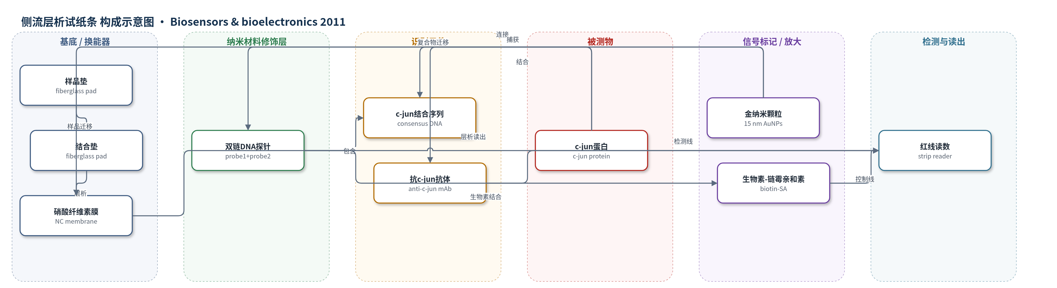
点击图片查看大图 · 依据论文自动绘制
传感器类型
侧流层析试纸条
检测对象
c-jun 蛋白(c-jun protein,DNA-binding protein);样品基质:纯化重组 c-jun 蛋白、HeLa 细胞粗裂解液(crude cell lysate)
检测原理
该传感器利用 DNA 探针-金纳米颗粒复合物识别活性 c-jun。15 nm AuNP 通过硫醇端连接含 c-jun 结合共识序列的 probe 1,并与生物素化 probe 2 杂交形成双链探针。样品中活性 c-jun 与双链 DNA 探针上的结合序列特异性结合,形成 AuNP-DNA-c-jun 复合物;未结合 c-jun 的探针-AuNP 复合物保持游离。复合物随层析液迁移至硝酸纤维素膜,检测区抗 c-jun 单克隆抗体捕获 c-jun,使 AuNP 在检测线聚集,产生红色检测线;剩余生物素化探针-AuNP 复合物被控制区链霉亲和素捕获形成控制线。检测线峰面积/光密度随 c-jun 活性增加而增强,因此可半定量反映转录因子活性。该法无需核酸酶、荧光或放射性标记,且仅检测能结合 DNA 的活性蛋白。
检测灵敏度
LOD: 0.2 footprint unit (fpu) c-jun(视觉检出阈值);细胞裂解液可见量: 100 μg total protein;检测时间: 10 min
效应效果
该传感器 10 min 内完成检测,视觉检出 0.2 fpu 纯化 c-jun,并可在 100 μg 以上 HeLa 粗裂解液中产生可见检测线。特异性方面,检测区无背景;突变结合序列试纸对 150 μg 裂解液无检测线;300 μg 卵白蛋白(OVA)无检测线,说明抗体可区分 c-jun 与非特异蛋白。热灭活 150 μg 裂解液无检测线,未处理 150 μg 裂解液、0.3 fpu 纯化 c-jun 及灭活裂解液加标 0.3 fpu 均产生检测线,后两者强度无显著差异(p<0.05,n=3),表明细胞成分不干扰且只检测活性 c-jun。与 EMSA 相比,EMSA 可检测 50 μg 裂解液,灵敏度更高,但本方法更快、无需电泳和标记,适合快速筛查转录因子活性。
传感器的构成
- 样品垫:玻璃纤维(fiberglass),浸样品垫缓冲液(1% Triton、1% BSA、2% 葡萄糖、50 mM 硼酸,pH 8.0),用于加样与样品释放。
- 结合垫:玻璃纤维(fiberglass),用于承载/释放 AuNP-DNA 探针复合物并启动层析。
- 硝酸纤维素膜(NC membrane):层析载体,检测区固定抗 c-jun 单克隆抗体(anti-c-jun mAb),控制区固定链霉亲和素(streptavidin, SA)。
- 金纳米颗粒(AuNPs):15 nm 金纳米颗粒,作为红色信号标记和 DNA 探针载体,经柠檬酸还原法制备。
- DNA 探针层:5′-巯基修饰 40-mer probe 1(SH-5′-AAAAAAAAAAAAAAAAAAAATCCGGCTGAATCATCAAGCG-3′)与 5′-生物素修饰 20-mer probe 2(biotin-5′-CGCTTGATGATTCAGCCGGA-3′)杂交形成双链探针,提供 c-jun 结合位点。
- 识别元件:双链 DNA 探针中的 c-jun 结合共识序列,特异性捕获活性 c-jun 蛋白。
- 捕获抗体:抗 c-jun 单克隆抗体(anti-c-jun mAb)固定于检测区,捕获 c-jun 并区分非特异蛋白。
- 控制捕获:链霉亲和素(SA)固定于控制区,通过 SA-生物素结合捕获剩余 probe-AuNP 复合物,指示试纸有效。
- 吸收垫:厚吸收纸(absorbent paper),吸收液体并维持层析流动。
中文摘要
本研究开发了一种基于免疫层析的侧流生物传感器,用于快速检测 DNA 结合蛋白。该传感器由样品垫、结合垫、硝酸纤维素膜和吸收垫四部分组成。含 c-jun 蛋白结合共识序列的 5′-巯基修饰 DNA 探针通过硫醇端固定在 15 nm 金纳米颗粒上,并与 5′-生物素修饰互补探针杂交形成双链 DNA 探针;抗 c-jun 单克隆抗体固定于硝酸纤维素膜检测区,链霉亲和素固定于控制区。样品中的 c-jun 蛋白与金纳米颗粒上的 DNA 结合序列结合,形成 AuNP-DNA-c-jun 复合物,经层析迁移后被检测区抗体捕获,形成红色检测线;剩余复合物由控制区链霉亲和素通过生物素-链霉亲和素作用捕获,形成控制线。该传感器可在 10 min 内视觉检测低至 0.2 footprint unit 的 c-jun,并成功用于 HeLa 细胞粗裂解液中 c-jun 活性检测,具有快速、简便、高特异性的优点。
英文摘要
A lateral flow biosensor based on an immuno-chromatographic assay has been developed for the detection of DNA-binding proteins. The biosensor is composed of four parts: a sample pad, a conjugate pad, a strip of nitrocellulose membrane and an absorbent pad. A DNA probe containing a specific protein binding consensus sequence is coated onto gold nanoparticles, while an antibody against the DNA-binding protein is immobilized onto a test zone of the nitrocellulose membrane. The target protein binds to the protein binding DNA sequence that is coated on the gold nanoparticles to form nanoparticle-DNA-protein complexes, and the complexes are then captured by the antibody immobilized on the test zone to form a red line for visual detection of the target protein. This biosensor was successfully applied to a DNA-binding protein, c-jun, and the developed biosensor allows for the rapid detection of down to 0.2 footprint unit of c-jun protein within 10 min. This biosensor was verified using HeLa cells and it visually detected c-jun activity in 100 μg of crude cell lysate protein. The antibody against c-jun used in the biosensor can distinguish c-jun from other nonspecific proteins, with high specificity.