组成图示
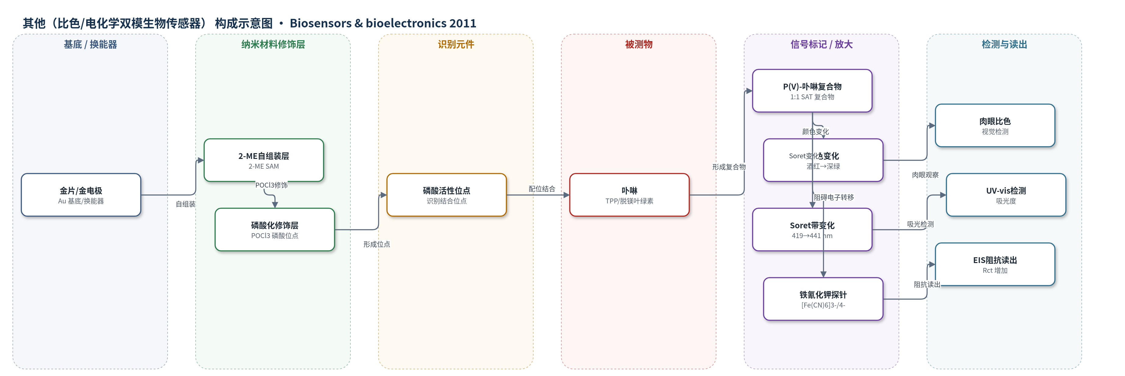
点击图片查看大图 · 依据论文自动绘制
传感器类型
其他(比色/电化学双模生物传感器)
检测对象
卟啉(porphyrin)、四苯基卟啉(TPP)、脱镁叶绿素(pheophytin);样品基质为二氯甲烷溶液及菠菜叶提取液
检测原理
该传感器基于磷酸基团与卟啉的特异性配位识别。金表面先自组装2-ME形成含羟基SAM,再用POCl3修饰生成磷酸活性位点。卟啉结合后形成1:1阳离子sitting-atop(SAT)P(V)-porphyrin复合物。比色信号来自卟啉平面畸变和共轭体系变化,Soret带由419 nm红移至441 nm,溶液由酒红变深绿,浓度越高颜色变化越明显直至位点饱和。电化学信号来自复合物在电极界面阻碍[Fe(CN)6]3-/4-电子转移,使电荷转移电阻Rct增大;Rct与TPP浓度对数在1.0×10−7 M至5.0×10⁻⁵ M范围内线性相关。
检测灵敏度
LOD: 比色法 1.0 × 10⁻⁶ M;EIS 3.0 × 10⁻⁸ M;线性范围: 1.0 × 10⁻⁷ M–5.0 × 10⁻⁵ M(Rct 对 TPP 浓度对数线性);R = 0.9984
效应效果
该传感器响应快,比色法3 s内可肉眼观察到卟啉溶液由酒红变深绿;修饰金片/电极在二氯甲烷溶液或4 ℃空气中保存数周后仍稳定。菠菜叶脱镁叶绿素加标回收率为91.7%、95.8%、105.6%和93.8%,RSD为1.98%–2.56%(n=4),与紫外分光光度法结果(0.20–0.23 μmol/L)一致。对胡萝卜素、类胡萝卜素、叶绿素和维生素无干扰。与传统光谱、HPLC、MS等方法相比,操作简便、成本低,可用于生物和医学体系中卟啉定量,并有望开发成类似pH试纸的变色产品。
传感器的构成
- 基底/换能器:金片或金电极(Au),提供导电基底并作为电化学阻抗换能器
- 自组装单分子层:2-巯基乙醇(2-ME)SAM,硫端锚定于金表面,暴露羟基(-OH)
- 磷酸化修饰层:三氯氧磷(POCl3)与羟基反应生成 -CH2-O-POCl2 和 (-CH2-O-)2POCl 磷酸活性位点
- 识别元件:表面磷酸基团,与卟啉配位形成 1:1 阳离子 sitting-atop(SAT)P(V)-porphyrin 复合物
- 光学信号元件:卟啉(TPP/脱镁叶绿素)自身颜色由酒红变深绿,Soret 带由 419 nm 红移至 441 nm
- 电化学探针:[Fe(CN)6]3-/4-(5.0 mM)与 0.1 M KCl,用于 EIS 监测电荷转移电阻 Rct
中文摘要
卟啉能够与磷特异性结合。本文报道了一种用于比色和电化学测定卟啉的新型双功能传感平台。将经2-巯基乙醇(2-ME)预处理的金片或金电极用三氯氧磷(POCl3)化学修饰,获得表面磷酸活性位点,并通过电化学阻抗谱(EIS)表征各修饰阶段。由于修饰金片/电极对卟啉的高亲和力,形成1:1阳离子sitting-atop(SAT)P(V)–卟啉复合物,使电极表面电子转移电阻增大。同时,肉眼可在3 s内观察到卟啉溶液由酒红色变为深绿色,UV–vis中Soret带也发生显著变化。两种传感体系对卟啉具有不同灵敏度:比色法检测限为1.0×10⁻⁶ M,EIS法检测限为3.0×10⁻⁸ M。该传感器已成功用于新鲜菠菜叶中脱镁叶绿素的测定,结果表明该双功能生物传感器操作简便、有效,适用于卟啉定量分析。
英文摘要
Porphyrin is able to specifically combine with phosphorus, thus a novel bifunctional sensing platform for determination of porphyrin by visual colorimetry and electrochemistry was demonstrated. A pretreated gold sheet (or electrode) with 2-mercatpoethanol (2-ME) was chemically modified by POCl(3) to obtain the surface phosphate active sites. The different stages of modified electrode were characterized by electrochemical impedance spectroscopy (EIS). The 1:1 cationic sitting-atop (SAT) complex P(V)-porphyrin was formed due to the high affinity of the modified gold sheet (or electrode) towards the porphyrin, resulting in electron transfer resistance increase of the electrode surface. Meanwhile, a dramatic color changing from burgundy to dark green of porphyrin solution was observed with the naked-eye within 3s. What's more, this was reflected by the notable change of the Soret band of porphyrin when using UV-vis. Two sensing systems provide different sensitivity for porphyrin analysis. With visual colorimetry, porphyrin can be detected at a level of 1.0×10(-6) M, whereas the detection limit of porphyrin is 3.0×10(-8) M using the EIS method. The practical application of the sensor to determination of pheophytin which was obtained from fresh spinach leaves has been accomplished. The results demonstrate the facility and effectivity of our introduced bifunctional biosensor for quantitative analysis of porphyrin.