组成图示
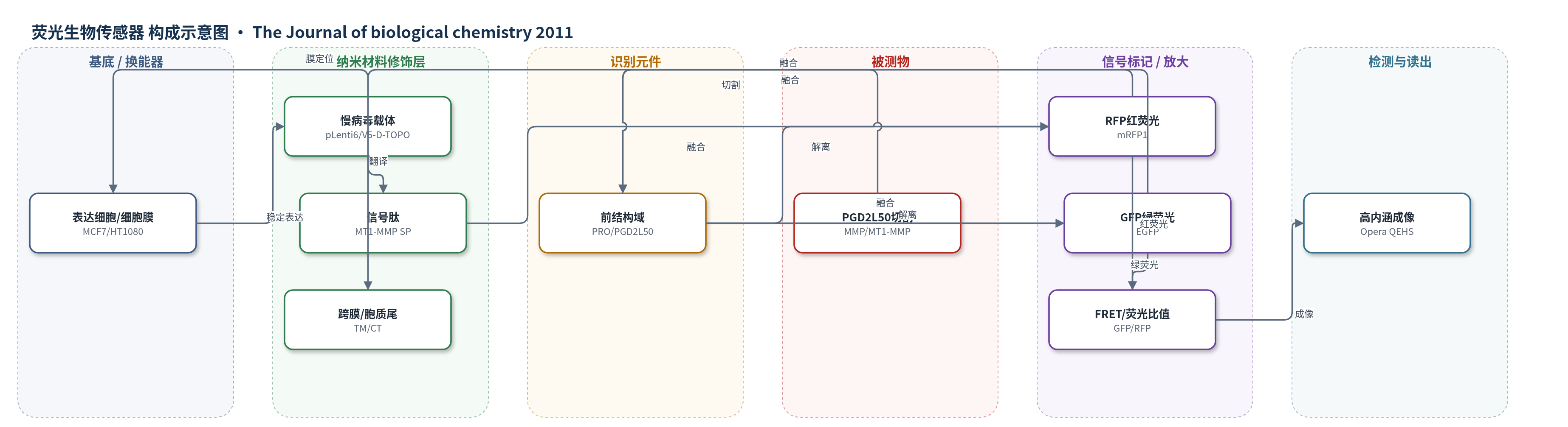
点击图片查看大图 · 依据论文自动绘制
传感器类型
荧光生物传感器
检测对象
MT1-MMP 前结构域 PGD2L50 内域切割事件(MT1-MMP prodomain PGD2L50 cleavage);样品基质:MCF7、HT1080、MDA-MB-435 培养细胞及小鼠原位乳腺癌异种移植肿瘤组织
检测原理
RFP-PRO-GFP 生物传感器由 MT1-MMP 信号肽、RFP、前结构域 PRO、GFP、跨膜域和胞质尾串联组成。完整传感器中,RFP 在 503 nm 处存在附加吸收峰,强烈猝灭/吸收 GFP 507 nm 发射,因此主要显示红荧光并可发生 FRET。当细胞内 MMP/MT1-MMP 在 PRO 的 PGD2L50 位点发生内域切割时,RFP 与 GFP 空间分离,GFP 绿荧光恢复,GFP/RFP 比值升高。L50D 突变使该切割位点失活,传感器保持完整,仅显示红荧光。因此信号变化反映前结构域切割事件或蛋白酶活性,而非直接定量外部分析物浓度。
检测灵敏度
原文未报告 LOD、线性范围、灵敏度斜率或相关系数。
效应效果
该荧光生物传感器在 HT1080、MCF7 和 MDA-MB-435 细胞中高尔基体中可被切割,而 L50D 突变传感器不被切割,仅呈红荧光,显示对 PGD2L50 切割位点具有特异性。表面生物素化证实完整与切割形式均可到达细胞表面,但切割主要发生在胞内。在小鼠原位乳腺癌异种移植中,MCF7-MT1 肿瘤 10 周体积比 MCF7-L50D 大 20 倍,L50D 肿瘤第 4 周约 25 mm3 后停止生长,E240A 无可检测肿瘤。L50D 肿瘤 Ki-67 降低,细胞周期基因显著下调,MMP-2 主要保持酶原状态,说明该传感器可反映 MT1-MMP 促肿瘤功能。
传感器的构成
- 表达载体/细胞基底:pLenti6/V5-D-TOPO 慢病毒载体与 MCF7、HT1080、MDA-MB-435 细胞,用于稳定表达膜定位生物传感器
- 信号肽层:MT1-MMP signal peptide (SP),引导生物传感器进入分泌途径并定位细胞膜
- 红色荧光报告层:monomeric red fluorescent protein 1 (RFP),完整传感器中产生红荧光并在切割后解离
- 识别/切割元件:MT1-MMP prodomain (PRO),含 PGD2L50 内域切割位点,被 MMP/MT1-MMP 切割
- 绿色荧光报告层:enhanced green fluorescent protein (GFP),融合于 PRO C 端 Val107,切割后绿荧光恢复
- 膜锚定层:MT1-MMP transmembrane domain (TM) 与 cytoplasmic tail (CT),将传感器锚定于细胞膜并参与转运
- 突变对照层:L50D 突变 PRO (RFP-L50D-GFP),失活 PGD2L50 切割位点,作为阴性对照
- 读出系统:Olympus BX51 荧光显微镜与 Opera QEHS 高内涵成像,检测 GFP、RFP、FRET 及 GFP/RFP 比值
中文摘要
侵袭性肿瘤细胞依赖细胞周围蛋白水解突破细胞外基质和基底膜屏障,膜型基质金属蛋白酶-1(MT1-MMP)是癌细胞表面蛋白水解事件的主要介质。MT1-MMP 以酶原形式合成,其 N 端抑制性前结构域维持潜伏状态;分泌途径中 furin 切割 R108RKR1112Y112 位点,但完整前结构域仍非共价抑制新生酶。作者构建含 MT1-MMP 前结构域的 RFP-PRO-GFP 荧光生物传感器及 PGD2L50 位点失活的 L50D 突变传感器,并在 MCF7、HT1080 和 MDA-MB-435 细胞中检测前结构域内域切割。结合稳定表达野生型、L50D 和 E240A MT1-MMP 的细胞、小鼠原位乳腺癌异种移植及全基因组转录谱分析,证明前结构域 PGD2L50 内域切割对 MT1-MMP 促肿瘤功能至关重要,并提示该多步失活机制可能普遍存在于 MMP 家族。
英文摘要
Invasive cancers use pericellular proteolysis to breach the extracellular matrix and basement membrane barriers and invade the surrounding tissue. Proinvasive membrane type-1 matrix metalloproteinase (MT1-MMP) is the primary mediator of proteolytic events on the cancer cell surface. MT1-MMP is synthesized as a zymogen. The latency of MT1-MMP is maintained by its N-terminal inhibitory prodomain. In the course of MT1-MMP activation, the R(108)RKR(111) ↓ Y(112) prodomain sequence is processed by furin. The intact prodomain released by furin alone, however, is a potent inhibitor of the emerging MT1-MMP enzyme. Evidence suggests that the prodomain undergoes intradomain cleavage at the PGD ↓ L(50) site followed by the release of the degraded prodomain by furin cleavage that finalizes the two-step activation event. These cleavages, only if combined, cause the activation of MT1-MMP. The significance of the intradomain cleavage in the protumorigenic program of MT1-MMP, however, remained unidentified. To identify this important parameter, in our current study, we used the cells that expressed the wild-type prodomain-based fluorescent biosensor and the mutant biosensor with the inactivated PGD↓L(50) cleavage site (L50D mutant) and also the cells with the enforced expression of the wild-type and L50D mutant MT1-MMP. Using cell-based tests, orthotopic breast cancer xenografts in mice, and genome-wide transcriptional profiling of cultured cells and tumor xenografts, we demonstrated that the intradomain cleavage of the PGD ↓ L(50) sequence of the prodomain is essential for the protumorigenic function of MT1-MMP. Our results emphasize the importance of the intradomain cleavages resulting in the inactivation of the respective inhibitory prodomains not only for MT1-MMP but also for other MMP family members.