组成图示
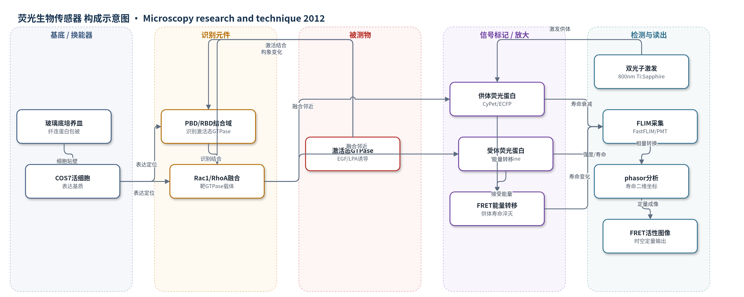
点击图片查看大图 · 依据论文自动绘制
传感器类型
荧光生物传感器
检测对象
Rac1 活性(Rac1 activity)、RhoA 活性(RhoA activity);样品基质:COS7 活细胞(EGF 或 LPA 刺激)
检测原理
该检测基于荧光蛋白 FRET 生物传感器。当 EGF 或 LPA 诱导 Rac1 或 RhoA 进入激活态时,PBD 或 RBD 识别并结合激活态 GTPase,使供体荧光蛋白(CyPet/ECFP)与受体荧光蛋白(YPet/Citrine)的距离或取向改变,发生非辐射偶极-偶极能量转移。FRET 使供体荧光强度下降、寿命缩短,受体发射增强。系统用 800 nm 双光子激光选择性激发供体,通过 FLIM 采集每个像素的供体衰减曲线,并用 phasor 变换将衰减映射为二维坐标。独立荧光团混合在 phasor 图中形成线性轨迹,而 FRET 引起的寿命变化形成曲线轨迹,因此可区分背景、未成熟蛋白和光漂白伪影,定量自由/结合或低/高 FRET 比例,实现活细胞内 GTPase 活性的时空成像。
检测灵敏度
原文未报告 LOD、线性范围、灵敏度斜率或相关系数。
效应效果
作者将 phasor-FLIM 与 GP 比率分析比较,表明 phasor 方法对双链/单链设计及细胞荧光伪影更稳健。Rac1 双链传感器未淬灭供体平均寿命 2.61 ns,FRET 态效率约 30%;RhoA 单链传感器残余低 FRET 效率为 4% 和 7%,高 FRET 效率为 35% 和 39%,说明单链存在基线 FRET,动态范围低于双链。强度法受供体/受体分布不均影响,在 Rac1 双链实验中将核周低供体浓度误判为 FRET;phasor-FLIM 给出与文献一致的边缘/后部突起定位。失活与组成活性对照均显示均匀低或高 FRET。作者认为该方法可用于传感器优化及多重荧光工具下的定量检测。
传感器的构成
- 基底/样品基质:35 mm 玻璃底培养皿,纤连蛋白包被,用于 COS7 细胞贴壁与成像
- 表达基质:COS7 活细胞,瞬时转染表达 GTPase 生物传感器,提供活细胞内检测环境
- 识别元件:PBD(PAK1 结合域)或 RBD(Rhotekin 结合域),特异性结合激活态 Rac1 或 RhoA
- 靶蛋白融合链:Rac1 或 RhoA 与荧光蛋白融合,作为被检测 GTPase 活性状态的结构载体
- 供体荧光蛋白:CyPet 或 ECFP,作为 FRET 供体,其荧光寿命在 FRET 发生时被淬灭
- 受体荧光蛋白:YPet 或 Citrine,作为 FRET 受体,接受供体能量并产生增强发射
- 构象/定位元件:单链设计中的 1L 连接序列及 kRas 脂质基序,调控 RhoA 膜定位与构象变化;双链设计为两条独立链,激活后结合
- 换能/读出系统:800 nm 双光子 Ti:Sapphire 激光激发供体,Zeiss LSM710 META 共聚焦显微镜、FastFLIM 与 PMT 采集供体/受体信号
中文摘要
本文提出基于荧光寿命成像显微镜(FLIM)的相量(phasor)方法用于生物传感器 Förster 共振能量转移(FRET)检测,并证明该方法对生物传感器设计以及细胞环境中固有的荧光伪影具有稳健性。作者以一系列双链和单链 GTPase 生物传感器为对象,这些传感器用于报告 Rac1 和 RhoA 活性的亚细胞定位;同时对所获得的 FLIM 数据采用广义极化(GP)方法进行同步比率 FRET 分析,以比较两种方法在时间和空间上定量成像生物传感器 FRET 信号的能力。结果表明,在相量图中,寿命分析将每种分子物种转换为二维坐标,使独立荧光团混合与由 FRET 引起的寿命变化得以区分。因此,可在图像每个像素中定量双链生物传感器的自由态与结合态,或单链生物传感器的低 FRET 与高 FRET 物种的比例。相量分析还能准确表征各传感器设计的固有物理性质,从而可用于开发阶段的传感器优化。作者认为,随着生物传感器更加复杂并与其他荧光分子工具多重化,基于 FLIM 相量方法的 FRET 检测将不仅成为其使用所必需,也将推动其发展。
英文摘要
We present here the phasor approach to biosensor Förster resonance energy transfer (FRET) detection by fluorescence lifetime imaging microscopy (FLIM) and show that this method of data representation is robust towards biosensor design as well as the fluorescence artifacts inherent to the cellular environment. We demonstrate this property on a series of dual and single chain biosensors, which report the localization of Rac1 and RhoA activity, whilst performing concomitant ratiometric FRET analysis on the acquired FLIM data by the generalized polarization (GP) approach. We then evaluate and compare the ability of these two methods to quantitatively image biosensor FRET signal as a function of time and space. We find that with lifetime analysis in the phasor plot each molecular species is transformed into a two-dimensional coordinate system where independent mixtures of fluorophores can be distinguished from changes in lifetime due to FRET. This enables the fractional contribution of the free and bound state of a dual chain biosensor or the low and high FRET species of a single chain biosensor to be quantified in each pixel of an image. The physical properties intrinsic to each biosensor design are also accurately characterized by the phasor analysis; thus, this method could be used to inform biosensor optimization at the developmental stage. We believe that as biosensors become more sophisticated and are multiplexed with other fluorescent molecular tools, biosensor FRET detection by the phasor approach to FLIM will not only become imperative to their use but also their advancement.