组成图示
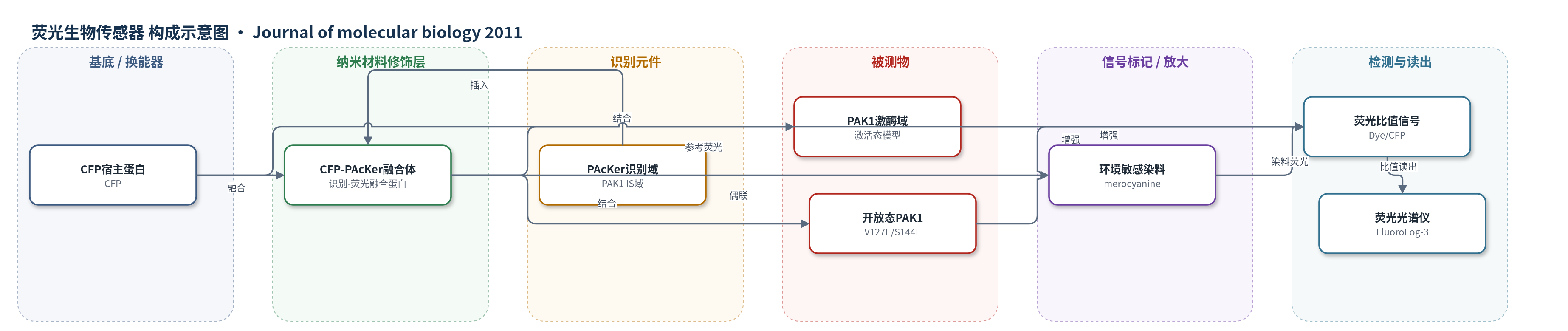
点击图片查看大图 · 依据论文自动绘制
传感器类型
荧光生物传感器
检测对象
PAK1激酶域(PAK1 kinase domain)、开放态全长PAK1(open full-length PAK1, PAK1V127E/S144E);样品基质:体外蛋白缓冲液,目标应用为活细胞内源PAK1
检测原理
CFP-PAcKer 中的 PAcKer 识别域特异性结合 PAK1 激酶域或开放态全长 PAK1 暴露的 IS 结合表面。结合事件使邻近结合界面的环境敏感 merocyanine 染料局部微环境改变,通常表现为溶剂可及性或极性降低,从而引起染料荧光增强。CFP 荧光作为内参,计算染料/CFP 荧光比值;该比值随 PAK1 激酶域或开放态 PAK1 浓度增加而上升,呈单点结合曲线。闭合态 PAK1 不暴露结合表面,因此不产生显著比值变化。该传感器无需酶促放大,依靠蛋白识别与染料环境响应实现激活态 PAK1 的荧光检测。
检测灵敏度
未报告 LOD、线性范围、灵敏度斜率或相关系数;报告结合常数:Kd: 400 nM(CFP-PAcKer 与 PAK1 激酶域,ITC);Kd: 3.3 μM(CFP-PAcKer 与 PAK1V127E/S144E);荧光比值拟合 Kd: 600 nM;荧光净增益: ∼40%。
效应效果
CFP-PAcKer 对 PAK1 激酶域的 ITC 亲和力为 400 nM,较野生型 IS 域(4 μM)提高约 10 倍;对闭合态全长 PAK1 无检测到的结合,对开放态 PAK1V127E/S144E 的 Kd 为 3.3 μM。PAcKer V127E 或 PAK1 L470E 突变均消除结合,说明识别界面正确。merocyanine 偶联效率约 100%;200 nM mero-CFP-PAcKer 滴定 PAK1 激酶域时,Dye/CFP 比值净增益约 40%,拟合 Kd 为 600 nM;PAK1 L470E 和闭合态 PAK1 无显著变化。1 μM GST-PAK5 仅引起小变化,>8 μM 时变化大于 PAK1,提示对 PAK5 结合较弱。作者认为该亲和试剂可用于活细胞内源 PAK1 激活动态成像。
传感器的构成
- 宿主荧光蛋白层:CFP(cyan fluorescent protein),提供稳定骨架与参考荧光(433 nm 激发/474 nm 发射)
- 识别元件层:PAcKer(重设计 PAK1 IS 域,插入 CFP 172-173 环区),特异性结合 PAK1 激酶域/开放态 PAK1
- 信号标记物层:merocyanine 染料(环境敏感荧光染料,偶联于 CFP-PAcKer L134C,结合后荧光增强,593 nm 激发/620 nm 发射)
- 靶标结合界面:PAK1 激酶域 C 叶/开放态 PAK1 的 IS 结合表面,与 PAcKer 互补结合
中文摘要
p21-激活激酶1(PAK1)的抑制开关(IS)域通过与激酶域结合,将全长 PAK1 稳定于非活性构象;小分子鸟苷三磷酸酶(GTPase)竞争性结合 IS 域可破坏自抑制并暴露激酶域表面的 IS 结合位点。为构建能选择性识别激活态 PAK1 的亲和试剂,作者利用分子建模对分离的 IS 域进行重新工程,使其可溶、稳定、不结合 GTPase,并更紧密地结合 PAK1 激酶域。研究测试了三种设计策略:前两种通过 N 端延伸与重设计扩大疏水核心;第三种将 N、C 端在空间上靠近,使该域可插入青色荧光蛋白(CFP)环区。最佳设计 CFP-PAcKer 基于第三种策略,以 400 nM 亲和力结合 PAK1 激酶域;对稳定于开放态的全长 PAK1 变体结合更强(Kd=3.3 μM),对闭合态全长 PAK1 无检测到的亲和力。通过在 CFP-PAcKer 结合位点邻近位置引入环境敏感荧光染料,可监测其与 PAK1 的结合。
英文摘要
The inhibitory switch (IS) domain of p21-activated kinase 1 (PAK1) stabilizes full-length PAK1 in an inactive conformation by binding to the PAK1 kinase domain. Competitive binding of small guanosine triphosphatases to the IS domain disrupts the autoinhibitory interactions and exposes the IS domain binding site on the surface of the kinase domain. To build an affinity reagent that selectively binds the activated state of PAK1, we used molecular modeling to reengineer the isolated IS domain so that it was soluble and stable, did not bind to guanosine triphosphatases and bound more tightly to the PAK1 kinase domain. Three design strategies were tested: in the first and second cases, extension and redesign of the N-terminus were used to expand the hydrophobic core of the domain, and in the third case, the termini were redesigned to be adjacent in space so that the domain could be stabilized by insertion into a loop in a host cyan fluorescent protein (CFP). The best-performing design, called CFP-PAcKer, was based on the third strategy and bound the kinase domain of PAK1 with an affinity of 400 nM. CFP-PAcKer binds more tightly to a full-length variant of PAK1 that is stabilized in the "open" state (K(d)=3.3 μM) than to full-length PAK1 in the "closed" state (undetectable affinity), and binding can be monitored with fluorescence by placing an environmentally sensitive fluorescence dye on CFP-PAcKer adjacent to the binding site.