组成图示
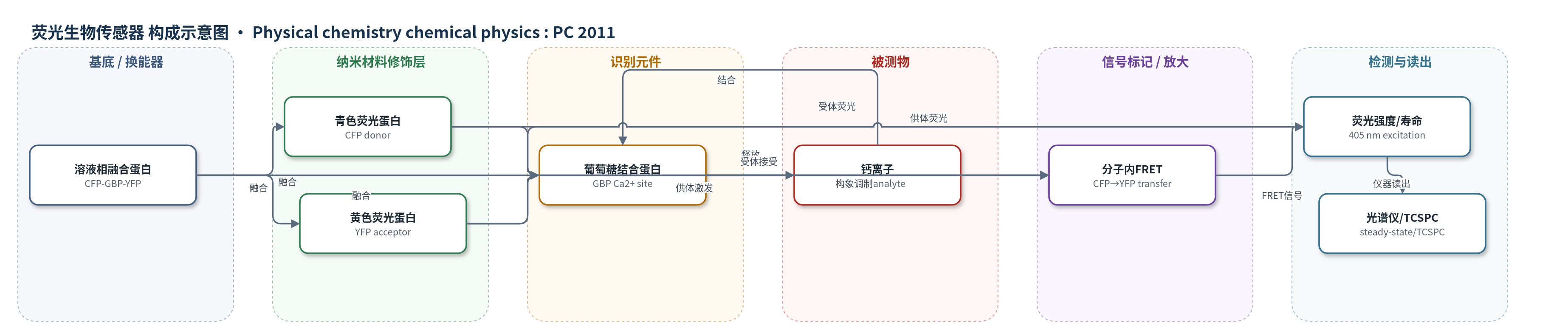
点击图片查看大图 · 依据论文自动绘制
传感器类型
荧光生物传感器
检测对象
钙离子(Ca2+);样品基质:磷酸盐缓冲液/溶液(phosphate saline),通过 CaCl2 或 EDTA 调节 Ca2+ 结合/释放状态。
检测原理
CFP–GBP–YFP 中,GBP 作为识别域,其 Ca2+ 结合位点被 Ca2+ 占据时发生闭合构象,使 CFP 与 YFP 距离缩短;EDTA 螯合 Ca2+ 后 GBP 开放,两荧光蛋白距离增大。405 nm 激发主要被 CFP 吸收,CFP 激发态能量通过 Förster 共振能量转移非辐射传递给 YFP,导致 CFP 荧光减弱、YFP 荧光增强。Ca2+ 结合分数越高,FRET 效率越高,YFP 发射越强,CFP 衰减越快,YFP 衰减出现能量馈入特征。通过稳态荧光强度和 TCSPC 荧光寿命可实时反映 Ca2+ 结合/释放引起的构象变化。
检测灵敏度
原文未报告 LOD、线性范围、灵敏度斜率或 R^2。
效应效果
论文未报告选择性、抗干扰、长期稳定性、RSD、实际样品加标回收率或与 ELISA/HPLC/qPCR 的对比。实验显示 Ca2+ 结合/释放过程可逆:EDTA 螯合 Ca2+ 后 FRET 降低,再加入 CaCl2 可恢复原始荧光谱形。稳态荧光与 TCSPC 寿命独立给出一致结果;EDTA 竞争释放的时间常数约 55 min。结合态与去钙态的聚合 FRET 效率分别约 0.25 和 0.05,对应 CFP–YFP 距离约 5.9 nm 和 8.0 nm;若按快/慢衰减分别计算,距离约 5.0–7.4 nm。作者认为该 FRET 蛋白可实时监测蛋白构象、开发生物传感器蛋白并表征蛋白–药物相互作用。
传感器的构成
- 基底/换能器:无固体基底,溶液相 CFP–GBP–YFP 融合蛋白作为光学换能器
- 识别元件:葡萄糖结合蛋白(GBP),含 Ca2+ 结合位点,结合/释放 Ca2+ 引起构象变化
- 供体荧光蛋白:青色荧光蛋白(CFP),吸收 405 nm 激发光并作为 FRET 供体
- 受体荧光蛋白:黄色荧光蛋白(YFP),接受 CFP 能量并增强 527 nm 荧光
- 信号标记/放大:CFP–YFP 分子内 FRET,Ca2+ 结合缩短 CFP–YFP 距离并提高 FRET 效率
- 样品介质:磷酸盐缓冲液(phosphate saline),可加入 CaCl2 或 EDTA 调节 Ca2+ 状态
- 信号读出:稳态荧光光谱与时间相关单光子计数(TCSPC)荧光寿命测量
中文摘要
本文报道了对一种生物合成融合蛋白 CFP–GBP–YFP 光学性质的系统表征。该蛋白由青色荧光蛋白(CFP)、葡萄糖结合蛋白(GBP)和黄色荧光蛋白(YFP)融合而成,其中 CFP 和 YFP 分别作为分子内荧光共振能量转移(FRET)的供体和受体。作者测量了融合蛋白的吸收、荧光发射、激发和荧光衰减,并与游离 CFP、YFP 比较,在光谱强度和荧光衰减中识别出能量转移特征。基于速率方程建立了描述荧光性质和能量转移的模型,并从实验数据中提取全部相关参数。融合蛋白结合钙离子后发生构象变化,导致 CFP 与 YFP 间能量转移效率改变。通过荧光强度和衰减测量追踪构象变化及钙结合反应动力学,并用速率方程模型解释结果。该蛋白构象变化的可视化有望作为实时研究蛋白结构变化、开发生物传感器蛋白以及表征蛋白–药物相互作用的分析工具。
英文摘要
This article reports the full characterisation of the optical properties of a biosynthesised protein consisting of fused cyan fluorescent protein, glucose binding protein and yellow fluorescent protein. The cyan and yellow fluorescent proteins act as donors and acceptors for intramolecular fluorescence resonance energy transfer. Absorption, fluorescence, excitation and fluorescence decays of the compound protein were measured and compared with those of free fluorescent proteins. Signatures of energy transfer were identified in the spectral intensities and fluorescence decays. A model describing the fluorescence properties including energy transfer in terms of rate equations is presented and all relevant parameters are extracted from the measurements. The compound protein changes conformation on binding with calcium ions. This is reflected in a change of energy transfer efficiency between the fluorescent proteins. We track the conformational change and the kinetics of the calcium binding reaction from fluorescence intensity and decay measurements and interpret the results in light of the rate equation model. This visualisation of change in protein conformation has the potential to serve as an analytical tool in the study of protein structure changes in real time, in the development of biosensor proteins and in characterizing protein-drug interactions.