组成图示
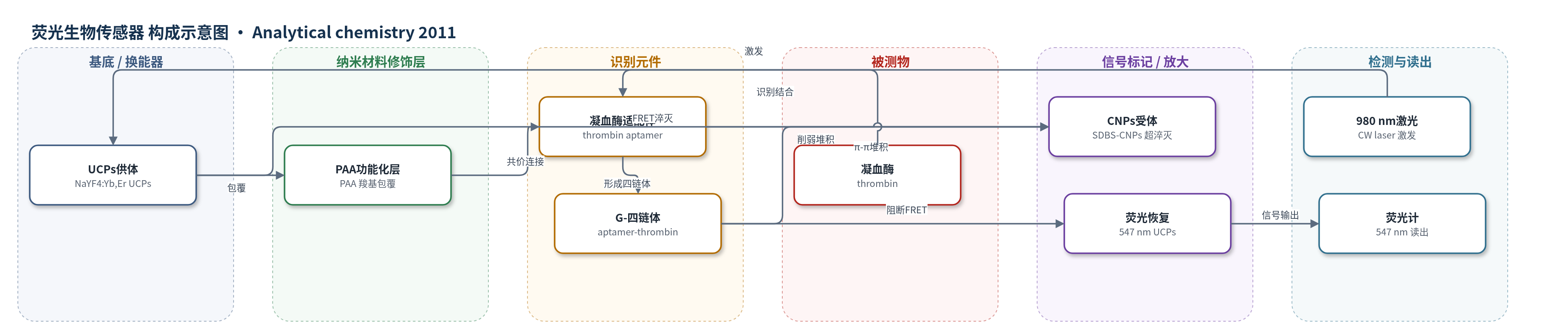
点击图片查看大图 · 依据论文自动绘制
传感器类型
荧光生物传感器
检测对象
凝血酶(thrombin);样品基质:水缓冲液、40倍稀释人血清、除纤维蛋白原后的人血浆
检测原理
PAA-UCPs 表面共价连接凝血酶适配体后,适配体通过 π-π 堆积吸附 SDBS-CNPs,使 UCPs 供体与 CNPs 受体接近,发生 FRET/表面能量转移,UCPs 547 nm 上转换荧光被 CNPs 超淬灭。加入凝血酶后,适配体与凝血酶特异性结合并形成 G-四链体,π-π 堆积减弱,CNPs 从 UCPs 表面分离,FRET 被阻断,UCPs 荧光随凝血酶浓度恢复。检测时用 980 nm 连续波激光激发 UCPs,读取 547 nm 发射强度;NIR 激发可避免生物基质自荧光和散射,UCPs 高发光与 CNPs 强淬灭共同提高信噪比。
检测灵敏度
LOD: 0.18 nM;线性范围: 0.5–20 nM;血清加标 LOD: 0.25 nM
效应效果
该传感器对凝血酶具有良好选择性:1 μM 金属离子、氨基酸和蛋白质等干扰物未引起明显荧光变化,而 20 nM 凝血酶可显著增强荧光。在 40 倍稀释人血清中,线性范围仍为 0.5–20 nM,检出限略升至 0.25 nM,说明抗复杂基质干扰能力较强。人血浆加标回收率为 96%–116%,RSD 约 4.2%–5.2%;三个血浆样品经 40 倍稀释校正后凝血酶浓度为 128、72 和 112 nM。与微流控量子点、时间分辨荧光及石墨烯 FRET 方法相比,该方法操作更简单、成本更低,并实现了血清和血浆中的实际定量,作者认为可用于凝血酶临床监测。
传感器的构成
- 荧光供体/换能纳米材料:NaYF4:Yb,Er 上转换磷光体(UCPs),980 nm 激发产生 547 nm 上转换荧光,作为 FRET 供体。
- 表面功能化层:聚丙烯酸(PAA)包覆 UCPs,提供水溶性及羧基反应位点,用于共价连接适配体。
- 识别元件:5'-NH2-GGTTGGTGTGGTTGG-3' 凝血酶适配体(thrombin aptamer),经 EDC/Sulfo-NHS 连接到 PAA-UCPs,识别凝血酶并诱导 G-四链体。
- 能量受体/淬灭层:SDBS 稳定碳纳米颗粒(CNPs),通过适配体 π-π 堆积非共价结合,超淬灭 UCPs 荧光。
- 封闭剂:Tris 封闭过量 Sulfo-NHS,终止偶联反应。
- 反应介质/样品基质:Tris-HCl 缓冲液(10 mM,150 mM NaCl,pH 7.4)、40 倍稀释人血清及除纤维蛋白原后的人血浆。
- 信号读出:980 nm 连续波激光激发,荧光光谱/光子计数系统记录 547 nm 发射强度。
中文摘要
本文报道了一种基于上转换磷光体(UCPs)向碳纳米颗粒(CNPs)荧光共振能量转移(FRET)的凝血酶适配体生物传感器。聚丙烯酸(PAA)功能化的 NaYF4:Yb,Er UCPs 通过 EDC/Sulfo-NHS 共价标记 5'-NH2-GGTTGGTGTGGTTGG-3' 凝血酶适配体,适配体再经 π-π 堆积吸附到 SDBS 稳定的 CNPs 表面,使供体与受体接近,UCPs 荧光被淬灭,优化条件下最大淬灭率达 89%。加入凝血酶后,适配体形成 G-四链体结构,π-π 作用减弱,CNPs 与 UCPs 分离,FRET 被阻断,UCPs 荧光随凝血酶浓度恢复,从而实现定量。该传感器在水缓冲液中凝血酶线性范围为 0.5–20 nM,检出限 0.18 nM;在人血清加标样品中获得相同线性范围,检出限 0.25 nM,并用于人血浆凝血酶水平监测,结果令人满意。这是首次将 UCPs 与 CNPs 作为供体-受体对构建 FRET 生物传感器。
英文摘要
We presented a new aptamer biosensor for thrombin in this work, which was based on fluorescence resonance energy transfer (FRET) from upconverting phosphors (UCPs) to carbon nanoparticles (CNPs). The poly(acrylic acid) (PAA) functionalized UCPs were covalently tagged with a thrombin aptamer (5'-NH(2)- GGTTGGTGTGGTTGG-3'), which bound to the surface of CNPs through π-π stacking interaction. As a result, the energy donor and acceptor were taken into close proximity, leading to the quenching of fluorescence of UCPs. A maximum fluorescence quenching rate of 89% was acquired under optimized conditions. In the presence of thrombin, which induced the aptamer to form quadruplex structure, the π-π interaction was weakened, and thus, the acceptor was separated from the donor blocking the FRET process. The fluorescence of UCPs was therefore restored in a thrombin concentration-dependent manner, which built the foundation of thrombin quantification. The sensor provided a linear range from 0.5 to 20 nM for thrombin with a detection limit of 0.18 nM in an aqueous buffer. The same linear range was obtained in spiked human serum samples with a slightly higher detection limit (0.25 nM), demonstrating high robustness of the sensor in a complex biological sample matrix. As a practical application, the sensor was used to monitor thrombin level in human plasma with satisfactory results obtained. This is the first time that UCPs and CNPs were employed as a donor-acceptor pair to construct FRET-based biosensors, which utilized both the photophysical merits of UCPs and the superquenching ability of CNPs and thus afforded favorable analytical performances. This work also opened the opportunity to develop biosensors for other targets using this UCPs-CNPs system.