组成图示
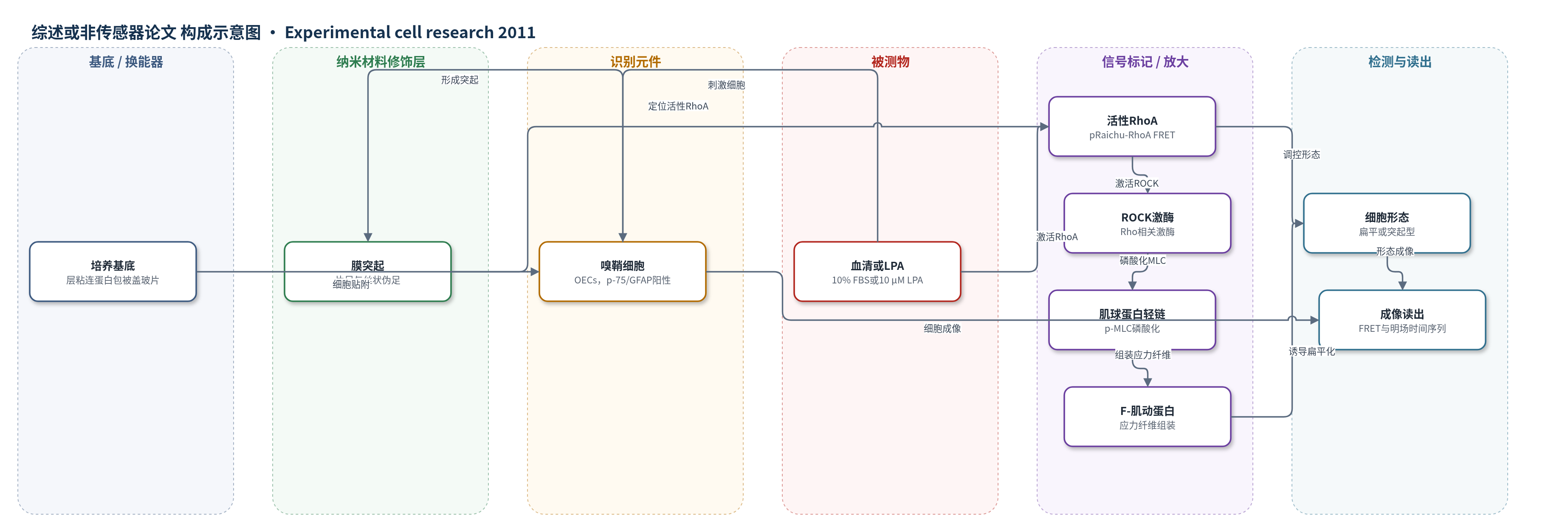
点击图片查看大图 · 依据论文自动绘制
传感器类型
综述或非传感器论文
检测对象
活性RhoA(active RhoA)、嗅鞘细胞形态(OEC morphology);样品基质:培养嗅鞘细胞(OECs)
检测原理
本文并非构建外部传感器,而是利用FRET探针pRaichu-RhoA在活细胞内监测RhoA活性。该探针由CFP标记的RhoA和YFP标记的Rhotekin RBD组成;当RhoA结合GTP并处于活性状态时,RBD与RhoA结合,使CFP与YFP距离缩短,产生FRET信号,FRET效率随活性RhoA水平升高而升高。血清或LPA刺激OECs后,RhoA被激活,FRET信号增强;活性RhoA进一步激活ROCK,ROCK磷酸化肌球蛋白轻链(MLC),激活肌球蛋白并促进F-肌动蛋白应力纤维组装,使细胞由突起型变为扁平型。抑制RhoA、ROCK或肌球蛋白则减少应力纤维,促进突起延长。信号通过三通道FRET成像和明场时间序列读出。
检测灵敏度
原文未报告LOD、线性范围、灵敏度斜率或相关系数。
效应效果
本文未评价传感器选择性、抗干扰、稳定性、重现性(RSD)或实际样品加标回收率,也未与ELISA、HPLC、qPCR等检测方法对比。实验表现方面,培养OECs纯度约95%;Y-27632梯度处理使19/20个扁平型OECs转变为突起型,PBS对照仅3/24;EGFP-WT-RhoA过表达使89.3±6.1%细胞呈扁平型,EGFP-DN-RhoA使84.4±16.6%呈突起型。Toxin-B、C3、Y-27632和Blebbistatin均剂量依赖阻断血清诱导的扁平化;LPA剂量依赖诱导扁平化且可被Y-27632阻断。作者认为该通路为调控OECs形态可塑性和神经再生相关表型提供了机制基础。
传感器的构成
- 识别元件:Rhotekin RBD(YFP–RBD),识别活性GTP-RhoA
- 信号标记物:CFP–RhoA与YFP–RBD融合蛋白pRaichu-RhoA,FRET报告RhoA活性
- 读出装置:Nikon Ti三通道荧光显微镜(CFP/FRET/YFP),输出FRET效率
中文摘要
嗅鞘细胞(OECs)是嗅觉系统中具有形态和功能可塑性的胶质细胞,培养条件下可呈现扁平型和突起型两种表型,且二者可相互转化,但调控其形态可塑性的分子机制尚不清楚。本研究利用RhoA荧光共振能量转移(FRET)生物传感器发现,活性RhoA主要分布于OECs的片足和/或丝状伪足,而非胞体;局部破坏这种活性RhoA分布可使细胞由扁平型转变为突起型,并促进突起生长。此外,RhoA通路抑制剂Toxin-B、C3、Y-27632或显性负性RhoA(DN-RhoA)过表达可阻断血清诱导的OECs由突起型向扁平型转变;而溶血磷脂酸(LPA)激活RhoA通路则促进突起型向扁平型转变。进一步结果表明,RhoA下游ROCK–肌球蛋白–F-肌动蛋白通路参与OECs形态可塑性。综上,RhoA–ROCK–肌球蛋白通路介导培养OECs对胞外信号的形态可塑性响应。
英文摘要
Olfactory ensheathing cells (OECs) are glial cells in the olfactory system with morphological and functional plasticity. Cultured OECs have the flattened and process-bearing shape. Reversible changes have been found between these two morphological phenotypes. However, the molecular mechanism underlying the regulation of their morphological plasticity remains elusive. Using RhoA FRET biosensor, we found that the active RhoA signal mainly distributed in the lamellipodia and/or filopodia of OECs. Local disruption of these active RhoA distributions led to the morphological change from the flattened into process-bearing shape and promoted process outgrowth. Furthermore, RhoA pathway inhibitors, Toxin-B, C3, Y-27632 or over-expression of DN-RhoA blocked serum-induced morphological change of OECs from the process-bearing into flattened shape, whereas the activation of RhoA pathway by lysophosphatidic acid (LPA) promoted the morphological change from the process-bearing into flattened shape. Finally, ROCK-Myosin-F-actin as a downstream of RhoA pathway was involved in morphological plasticity of OECs. Taken together, these results suggest that RhoA-ROCK-Myosin pathway mediates the morphological plasticity of cultured OECs in response to extracellular cues.