组成图示
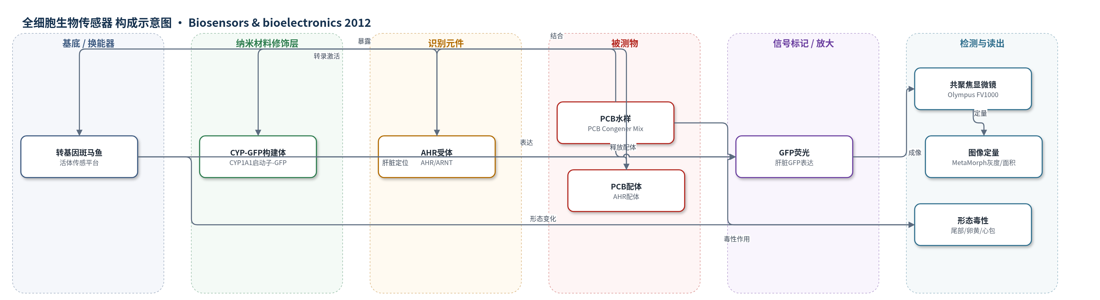
点击图片查看大图 · 依据论文自动绘制
传感器类型
全细胞生物传感器
检测对象
多氯联苯(polychlorinated biphenyls, PCBs);样品基质:淡水/水样(实验为PCB水溶液)
检测原理
PCBs作为芳香烃受体(AHR)配体进入转基因斑马鱼细胞后,与AHR结合,促使AHR转位入核并与ARNT形成二聚体,结合CYP1A1启动子区的异源物反应元件(XRE),从而诱导CYP1A1启动子驱动GFP转录。GFP在肝脏等组织积累,经488 nm激发产生绿色荧光。荧光平均灰度和表达面积随PCBs浓度变化:低浓度(0.02–0.08 μg/ml)诱导增强,0.08 μg/ml时强度最高,0.04 μg/ml时面积最大;高浓度(0.4–0.8 μg/ml)因肝损伤或表达耗竭而下降。时间上,GFP在暴露3 h后出现,5–6 h达最大后趋于平稳。该过程以报告基因表达实现生物放大和活体定位。
检测灵敏度
可检测最低浓度: 0.02 μg/ml;剂量响应关系: 0.2–0.8 μg/ml
效应效果
该活体传感器对PCBs呈浓度依赖响应:0.08 μg/ml时平均灰度由对照20.44±1.30升至43.02±0.14,增加110%;GFP面积在0.02 μg/ml最大,0.8 μg/ml降至1852±324 μm2(对照3996±427 μm2)。各浓度强度RSD为0.56%–33.48%,面积RSD为2.60%–30.34%(N=5)。暴露3 h出现GFP,5 h达最大,20 h稳定。与EROD、肝重和免疫组化相比,CYP-GFP可活体、实时、定位CYP1A1诱导,并同步观察尾部扭曲、卵黄扩张和心包水肿等毒性,无需牺牲鱼。作者认为其可监测低于LD50和德国TDI的淡水PCBs/PAHs污染。
传感器的构成
- 活体基底:转基因斑马鱼(transgenic zebrafish, Danio rerio),作为整体活体传感平台,肝脏为主要信号表达部位
- 遗传修饰层:CYP-GFP构建体(CYP1A1 promoter-GFP),由333 bp CYP1A1启动子融合GFP报告基因,用于响应PCBs诱导
- 表达载体:pZsGreen1-1载体(Clontech #632473),用于克隆CYP1A1启动子并驱动GFP表达
- 识别元件:芳香烃受体(AHR)及其核转位伴侣ARNT,结合PCBs后进入细胞核并激活CYP1A1启动子
- 信号标记物:绿色荧光蛋白(GFP),在CYP1A1启动子诱导下表达,提供可成像荧光信号
- 样品基质:双蒸馏水或PCB水溶液(PCB Congener Mix, Perkin Elmer),用于暴露转基因鱼
- 读出装置:Olympus FV1000共聚焦扫描显微镜/立体显微镜(488 nm激发)与MetaMorph软件,用于采集和定量GFP荧光
中文摘要
细胞色素P450(CYPs)在降解内源底物和解毒致癌物中具有重要作用,因此可作为评估水生环境中多环芳烃(PAHs)水平的生物标志物。本研究利用CYP1A1启动子驱动的绿色荧光蛋白(CYP-GFP)构建体,建立了转基因斑马鱼品系,用于检测多氯联苯(PCBs)毒性。实验采用0.02、0.04、0.08、0.4和0.8 μg/ml的PCBs处理转基因鱼。对照鱼肝脏仅显示低强度荧光;暴露PCBs后,鱼出现卵黄扩张、尾部扭曲和心包区膨胀等形态变化。绿色荧光信号随PCBs浓度和暴露时间变化:0.08 μg/ml处理时荧光强度最高,0.04 μg/ml时荧光面积最大。GFP在暴露3 h后开始表达,6 h时强度增加并趋于平稳。由于GFP表达响应迅速且对低浓度PAHs敏感,该转基因斑马鱼可作为活体成像和监测水污染的有力工具。
英文摘要
Cytochrome P450 (CYPs) is significant in degradation of endogenous substrates and detoxification of carcinogens, therefore it is a biomarker for assessment of polycyclic aromatic hydrocarbons (PAHs) level in aquatic environment. In the present study, a transgenic line of zebrafish had been generated using a CYP-green fluorescence protein (CYP-GFP) construct, driven by CYP1A1 promoter. Polychlorinated biphenyls (PCBs) were used as toxicant, in concentrations of 0.02 μg/ml, 0.04 μg/ml, 0.08 μg/ml, 0.4 μg/ml, and 0.8 μg/ml. The transgenic control fish showed low intensity of fluorescence in the liver. After exposed to PCBs, zebrafish had morphological changes such as expansion of yolk, contortion of tails and inflation of pericardial area. Green fluorescence signals were found to express according to concentrations and time. The green fluorescence signal was most intense after treatment with 0.08 μg/ml PCBs. However, the maximum area of green fluorescent signal was found at 0.04 μg/ml PCBs. GFP started to express at 3h exposure to PCBs, increasing its intensity until 6 h exposure, and then level off. Since the GFP expression is fast responding and is sensitive to low PAHs concentrations, transgenic fish is a good tool for live imaging and monitoring of aquatic contamination.