组成图示
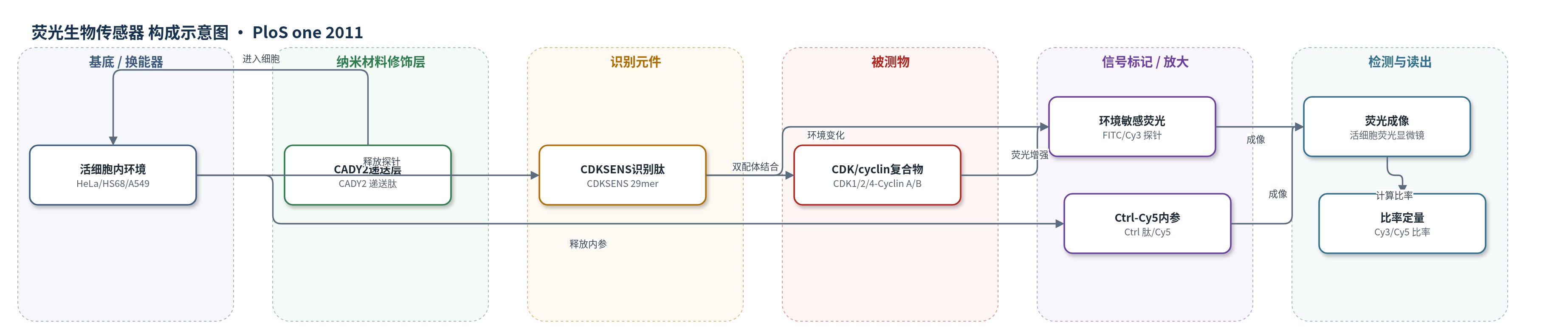
点击图片查看大图 · 依据论文自动绘制
传感器类型
荧光生物传感器
检测对象
CDK/周期蛋白复合物(CDK/cyclin complexes,含 CDK1/CDK2/CDK4 与 Cyclin A/B/D/E);样品基质:活细胞(HeLa、HS68、U20S、MCF-7、A549、HT2-19)及细胞裂解液。
检测原理
CDKSENS 为双配体荧光肽,一端含 CDK 底物结合序列 HHAGPRK,另一端含周期蛋白 RXL 招募序列 RRLFGE,可协同识别 CDK 催化区与周期蛋白沟槽,优先结合 CDK/cyclin 异二聚体而非单体。结合后,偶联在肽链内部的 FITC 或 Cy3 环境敏感探针从水相进入复合物低极性微环境,发生溶剂致变色,荧光强度增强约 3–4 倍。活细胞中,CADY2 以非共价复合物形式同时递送 CDKSENS-Cy3 与 Ctrl-Cy5;CDKSENS-Cy3 荧光随内源 CDK/cyclin 复合物丰度升高而增强,Ctrl-Cy5 作为内参校正递送、成像和细胞形态差异。最终通过 Cy3/Cy5 比率实现标准化定量,无需酶促放大,信号放大主要来自双配体协同识别和探针环境敏感性。
检测灵敏度
原文未报告 LOD、线性范围、灵敏度斜率或相关系数。
效应效果
体外滴定显示 CDKSENS 对 CDK2/CyclinA、CDK1/CyclinB、CDK2/CyclinB、CDK1/CyclinA 复合物 Kd 为 8±5、6±2、9±4、18±6 nM,对单体亲和力较低,对 GST/MBP 无结合,选择性良好。细胞裂解液 pulldown 可保留内源复合物,不识别单体 CDK。活细胞比率定量中,CDK1 降低约 80% 时比率下降 20%;CDK2 降低 50% 时下降 20%;CyclinB 降低 36% 时下降 15%。HeLa 与 HS68 平均差 15%,A549/MCF7 比 HeLa 低 18–20%,U20S 与 HeLa 无显著差异,与 Western blot 一致。重复实验标准偏差低,方法非侵入、无需固定或提取,可用于治疗监测、癌症诊断和药物发现。
传感器的构成
- 识别元件:CDKSENS 29mer 肽,含 CDK 结合序列 HHAGPRK、周期蛋白结合序列 RRLFGE 及 linker RVHER-YCSPTAGSAK,通过双配体识别 CDK/cyclin 复合物
- 信号标记物:FITC 或 Cy3 环境敏感荧光探针,偶联于 CDKSENS 内部半胱氨酸,结合靶标后因溶剂极性变化而荧光增强
- 比率内参:Ctrl 肽(VESSDTIDNVKSKIQDKEGC)标记 Cy5,作为内参用于 CDKSENS-Cy3/Ctrl-Cy5 比率定量
- 递送元件:细胞穿透肽 CADY2,与 CDKSENS-Cy3 和 Ctrl-Cy5 形成非共价复合物,介导进入活细胞
- 体外捕获基质:CNBr Sepharose 琼脂糖微球,偶联 CDKSENS 或 Ctrl 肽,用于从细胞裂解液中 pulldown 捕获 CDK/cyclin 复合物
- 读出系统:荧光显微镜或 Polarstar 荧光分光光度计,测量 Cy3/Cy5 或 FITC 荧光强度
中文摘要
细胞周期依赖性激酶(CDK)在协调细胞生长与分裂及维持癌细胞增殖中起核心作用,是重要药物靶点。然而,目前缺乏直接评估活细胞中 CDK 相对丰度的方法,现有手段主要依赖固定细胞的抗原检测和蛋白质组分析。为直接探测这些激酶,作者开发了一种荧光肽生物传感器 CDKSENS。该传感器在体外对 CDK 和周期蛋白具有双配体亲和力,可保留细胞裂解液中的内源 CDK/周期蛋白复合物,并带有环境敏感荧光探针,识别靶标后荧光显著增强。CDKSENS 通过与细胞穿透载体 CADY2 形成复合物进入活细胞,利用荧光成像和比率定量评估 CDK/周期蛋白相对丰度。该技术可直接、敏感地读出 CDK/周期蛋白复合物水平,报告干扰单个 CDK 或周期蛋白时复合物形成的差异,并能区分不同健康与癌细胞系中复合物表达高低。它以非侵入方式提供单个亚基抗原检测无法获得的复合物整体状态信息,无需细胞固定或提取,为监测 CDK/周期蛋白相关治疗反应、基于细胞的药物发现和荧光癌症诊断提供前景。
英文摘要
Cyclin-dependant kinases play a central role in coordinating cell growth and division, and in sustaining proliferation of cancer cells, thereby constituting attractive pharmacological targets. However, there are no direct means of assessing their relative abundance in living cells, current approaches being limited to antigenic and proteomic analysis of fixed cells. In order to probe the relative abundance of these kinases directly in living cells, we have developed a fluorescent peptide biosensor with biligand affinity for CDKs and cyclins in vitro, that retains endogenous CDK/cyclin complexes from cell extracts, and that bears an environmentally-sensitive probe, whose fluorescence increases in a sensitive fashion upon recognition of its targets. CDKSENS was introduced into living cells, through complexation with the cell-penetrating carrier CADY2 and applied to assess the relative abundance of CDK/Cyclins through fluorescence imaging and ratiometric quantification. This peptide biosensor technology affords direct and sensitive readout of CDK/cyclin complex levels, and reports on differences in complex formation when tampering with a single CDK or cyclin. CDKSENS further allows for detection of differences between different healthy and cancer cell lines, thereby enabling to distinguish cells that express high levels of these heterodimeric kinases, from cells that present decreased or defective assemblies. This fluorescent biosensor technology provides information on the overall status of CDK/Cyclin complexes which cannot be obtained through antigenic detection of individual subunits, in a non-invasive fashion which does not require cell fixation or extraction procedures. As such it provides promising perspectives for monitoring the response to therapeutics that affect CDK/Cyclin abundance, for cell-based drug discovery strategies and fluorescence-based cancer diagnostics.