组成图示
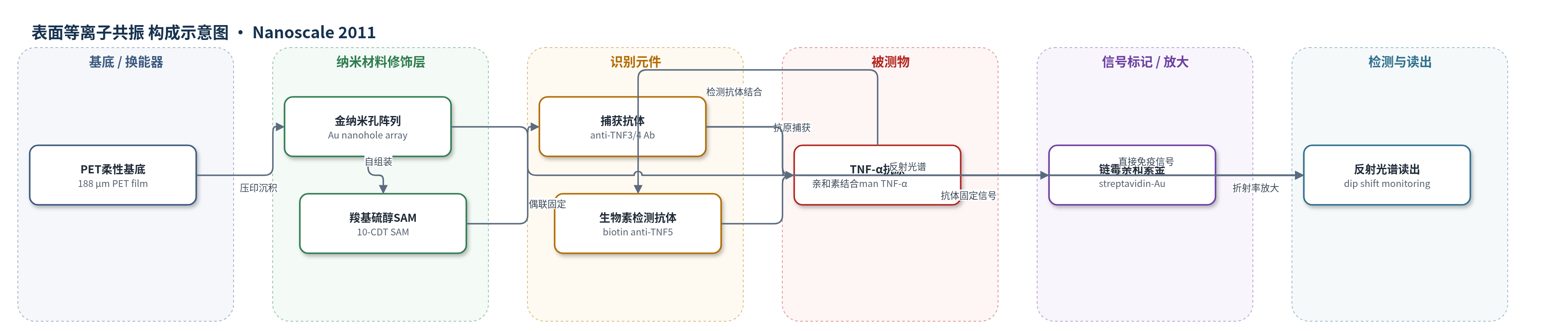
点击图片查看大图 · 依据论文自动绘制
传感器类型
表面等离子共振(SPR)生物传感器
检测对象
肿瘤坏死因子-α(TNF-α,tumor necrosis factor-alpha);样品基质:PBS 缓冲液(水相样品)
检测原理
该传感器基于金纳米孔阵列的等离激元共振。垂直入射光与金/介质界面自由电子耦合,激发 Bloch 波表面等离激元极化激元(SPP-BW)及孔局域表面等离激元(LSPR),在反射光谱中形成共振谷。当 TNF-α 与固定于 10-CDT 自组装单分子层上的捕获抗体结合后,界面局部折射率和质量增加,共振谷向长波方向移动。随后生物素化检测抗体结合抗原,链霉亲和素-金纳米颗粒通过生物素-亲和素作用进一步沉积在表面,形成非酶信号放大。反射光谱仪监测共振谷波长位移,其大小随 TNF-α 浓度增加而增大,从而实现定量检测。
检测灵敏度
LOD: 21 ng mL−1(streptavidin-gold 放大);LOD: 45 ng mL−1(direct immunoassay);灵敏度斜率: 495.0 nm RIU−1(periodicity 600 nm);FOM: 20.2;R^2 = 0.9998(bulk refractive index);S = 0.9889d − 94.58(R^2 = 0.9995)
效应效果
该器件采用金纳米孔阵列,对氧化稳定;压印模具重复使用100次以上仍保持均匀结构,可重复性良好。折射率测量中,1.000–1.700范围内共振谷位移与折射率线性相关,R^2=0.9998,清洗后信号可回到初始点。TNF-α检测中,链霉亲和素-金放大后LOD为21 ng/mL,直接免疫为45 ng/mL,较无标记Flexchip的120 ng/mL IgG约提高3倍。灵敏度495.0 nm RIU−1约为金胶体66.5 nm RIU−1的7倍,FOM 20.2高于银纳米立方5.4和NSL纳米井14.5。作者认为该低成本、可见光、可量产芯片适合POCT平台。
传感器的构成
- 基底:PET 薄膜(厚 188 μm),经 UV 纳米压印形成圆形纳米孔阵列,作为柔性光学基底
- 等离激元换能层:底部填充金纳米孔阵列(Au nanohole array,金厚 100 nm、孔深 50 nm、周期 600 nm、孔径 300 nm),激发 SPP-BW/LSPR 并产生反射光谱 dip
- 自组装单分子层:10-羧基癸硫醇(10-CDT)在 Au 表面形成 SAM,提供羧基用于抗体偶联
- 识别元件:抗 TNF-α 捕获抗体(mouse anti-TNF3/4)经 EDC/NHS 偶联固定,特异性结合 TNF-α
- 封闭剂:BSA(牛血清白蛋白)封闭未结合位点,降低非特异吸附
- 检测抗体:生物素化抗 TNF-α 抗体(biotinylated anti-TNF5)结合抗原,引入生物素信号位点
- 信号标记/放大:链霉亲和素-金纳米颗粒(streptavidin-gold,40 nm Au)通过生物素-亲和素结合,增加界面质量/折射率并放大 dip shift
- 微流控与读出:PDMS 微通道(2 mm×10 mm×20 mm)覆盖传感区,配合卤素光源、光纤束和分光光度计监测反射光谱 dip 移动
中文摘要
本文报道了一种可大规模生产的芯片式等离激元纳米孔阵列生物传感器。该器件以紫外固化聚氨酯丙烯酸酯(PUA)经纳米压印形成圆形纳米孔阵列,再在聚对苯二甲酸乙二醇酯(PET)薄膜上电子束沉积金薄膜,构成底部填充金纳米孔阵列。通过优化金膜厚度、孔深和孔周期,可获得最大反射光谱谷移,该谷移对应器件表面对折射率变化的灵敏度。作者利用三维时域有限差分(FDTD)方法计算反射光谱,并与实验结果一致。将纳米孔周期调节至400–900 nm,使共振谷位于可见光区,便于测量水相样品并减少水吸收干扰。优化后的器件被封装在聚二甲基硅氧烷(PDMS)微流控通道中,通道宽2 mm、深20 mm。作为概念验证,该传感器通过直接免疫反应检测肿瘤坏死因子-α(TNF-α),并采用胶体金标记代替酶放大,获得21 ng/mL的检测限。
英文摘要
We have developed a polymer film based plasmonic device whose optical properties are tuned for measuring biological samples. The device has a circular nanohole array structure fabricated with a nanoimprint technique using a UV curable polymer, and then gold thin film is deposited by electron beam deposition. Therefore, the device is mass-producible, which is also very important for bioaffinity sensors. First the gold film thickness and hole depth were optimized to obtain the maximum dip shift for the reflection spectra. The dip shift is equivalent to the sensitivity to refractive index changes at the plasmonic device surface. We also calculated the variation in reflection spectra by changing the above conditions using the finite-difference time domain method, and we obtained agreement between the theoretical and experimental curves. The nanohole periodicity was adjusted from 400 to 900 nm to make it possible to perform measurements in the visible wavelength region to measure the aqueous samples with less optical absorption. The tuned bottom filled gold nanohole array was incorporated in a microfluidic device covered with a PDMS based microchannel that was 2 mm wide and 20 μm deep. As a proof of concept, the device was used to detect TNF-α by employing a direct immunochemical reaction on the plasmonic array, and a detection limit of 21 ng mL(-1) was obtained by amplification with colloidal gold labeling instead of enzymatic amplification.