组成图示
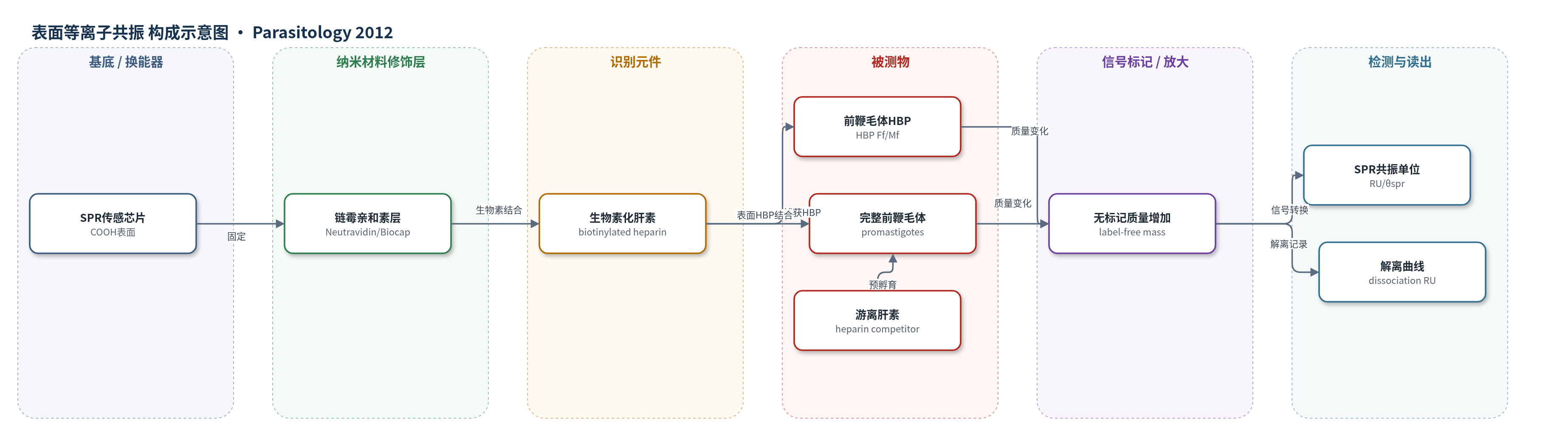
点击图片查看大图 · 依据论文自动绘制
传感器类型
表面等离子共振(SPR)生物传感器
检测对象
巴西利什曼原虫前鞭毛体肝素结合蛋白(heparin-binding proteins, HBPs;HBP Ff/HBP Mf),样品基质:鞭毛/膜亚细胞蛋白组分及完整前鞭毛体PBS悬浮液
检测原理
SPR芯片的羧基表面先固定链霉亲和素,再固定生物素化肝素,形成肝素识别界面。当含HBP的前鞭毛体或HBP Ff/HBP Mf蛋白流过芯片时,HBP与固定化肝素特异性结合,使界面质量增加,引起表面等离子共振角θspr变化,并以共振单位RU实时记录结合与解离。结合量随HBP浓度或细胞数增加而升高;游离肝素预孵育可占据HBP结合位点,降低RU,验证结合特异性。该法为无标记直接质量传感,不依赖酶或荧光放大,可实时获得结合/解离动力学。
检测灵敏度
未报告LOD、线性范围、灵敏度斜率或相关系数。
效应效果
SPR证实前鞭毛体表面存在HBP:结合/解离RU为33.0±2.0和14±1.0,较肝素基线高1.6倍。游离肝素预孵育呈剂量依赖性抑制,1.0、0.1、0.01 μg/ml分别抑制48%、39%、33%。HBP Ff解离RU为445±30(约0.4 ng/mm2),HBP Mf为175±20(约0.1 ng/mm2),分别较基线高53倍和20倍;BSA阴性对照RU仅2.0±0.3、1.2±0.1、0.4±0.3。肝素固定化KD=320 nM。作者认为SPR可实时无标记检测HBP,支持其参与寄生虫-媒介相互作用。
传感器的构成
- 基底/换能器:SPR传感芯片(COOH表面,SensiQ Pioneer),提供表面等离子共振光学换能
- 固定化层:Neutravidin(链霉亲和素,Biocap涂层),固定于羧基芯片表面,用于捕获生物素化肝素
- 识别/配体层:生物素化肝素(biotinylated heparin,0.5 μg),固定于链霉亲和素,作为肝素类似物识别HBP
- 信号层:无标记质量传感,HBP结合引起界面质量增加,不依赖荧光/酶标记
- 缓冲介质:PBS(磷酸盐缓冲液),维持结合条件并冲洗非特异结合
- 竞争抑制试剂:游离肝素(1.0、0.1、0.01 μg/ml),预孵育寄生虫以竞争HBP结合位点
- 信号读出:SPR共振角变化(θspr)以共振单位(RU)实时记录结合与解离
中文摘要
利什曼病是由媒介传播的重要公共卫生问题,利什曼原虫中的糖胺聚糖配体可作为控制该病的新靶点。本研究报道了巴西利什曼原虫(Leishmania (Viannia) braziliensis)前鞭毛体中肝素结合蛋白(HBPs)的亚细胞分布及其生化特性。前鞭毛体经差速离心分离为鞭毛和膜组分,并分别上样HiTrap肝素亲和层析柱,洗脱的肝素结合组分分别命名为HBP Ff和HBP Mf。HBP Ff中HBP浓度高于HBP Mf,SDS-PAGE显示两组分均含约65和55 kDa两条主要蛋白带。65 kDa蛋白具有明胶酶活性,并可被1,10-菲啰啉抑制。通过表面等离子共振(SPR)生物传感器分析,将前鞭毛体与肝素包被传感芯片结合,证实HBP定位于前鞭毛体表面;用不同浓度肝素预孵育寄生虫后,结合呈剂量依赖性抑制,提示两组分具有不同的肝素结合能力。结论表明,鞭毛或膜来源的蛋白组分具有金属蛋白酶特征并能结合糖胺聚糖。
英文摘要
Leishmaniasis is a vector-borne disease and an important public health issue. Glycosaminoglycan ligands in Leishmania parasites are potential targets for new strategies to control this disease. We report the subcellular distribution of heparin-binding proteins (HBPs) in Leishmania (Viannia) braziliensis and specific biochemical characteristics of L. (V.) braziliensis HBPs. Promastigotes were fractionated, and flagella and membrane samples were applied to HiTrap Heparin affinity chromatography columns. Heparin-bound fractions from flagella and membrane samples were designated HBP Ff and HBP Mf, respectively. Fraction HBP Ff presented a higher concentration of HBPs relative to HBP Mf, and SDS-PAGE analyses showed 2 major protein bands in both fractions (65 and 55 kDa). The 65 kDa band showed gelatinolytic activity and was sensitive to inhibition by 1,10-phenanthroline. The localization of HBPs on the promastigote surfaces was confirmed using surface plasmon resonance (SPR) biosensor analysis by binding the parasites to a heparin-coated sensor chip; that was inhibited in a dose-dependent manner by pre-incubating the parasites with variable concentrations of heparin, thus indicating distinct heparin-binding capacities for the two fractions. In conclusion, protein fractions isolated from either the flagella or membranes of L. (V.) braziliensis promastigotes have characteristics of metallo-proteinases and are able to bind to glycosaminoglycans.